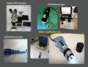
Different tools for nailfold capillaroscopic analysis.
Interest in viewing the nail capillaries dates to the late 17th century. Later research by Maurice Raynaud and others in the late 19th and early 20th century first established a direct link between the nailfold capillaries and certain medical conditions. Although underutilized in the past, with the advent of modern digital equipment and the validation of evidence-based methodologies, this is changing. Capillaroscopy is set to become a more important tool for rheumatologists, especially in the evaluation of Raynaud’s phenomenon and systemic sclerosis.1,2
Nail Capillary Anatomy
Capillaroscopy is a method used to examine a patient’s microcirculation and assess pathological changes. It is completely safe and noninvasive. Using some sort of microscopy, the clinician looks through the epidermis of the nailfold to view the capillaries of the distal papillae.3
Ariane Herrick, MD, is professor of rheumatology at the University of Manchester and honorary consultant rheumatologist at Salford Royal NHS Foundation Trust, England. She explains that nailfold anatomy makes it an ideal place to evaluate the microcirculation. In most of the skin, capillaries run perpendicular to the skin surface, and only the tip of the loop is visible.
Dr. Herrick explains, “At the nailfold, capillaries run parallel, rather than perpendicular, to the skin surface, and they can, therefore, be visualized noninvasively when magnified. Capillaries themselves are invisible, but what can be seen is the column of red blood cells within the capillaries.”
This is of interest to rheumatologists in evaluating systemic diseases that involve vascular damage. In some cases, pathogenic changes in capillary morphology may long predate the onset of clinical symptoms. And in patients already diagnosed with a systemic disease, such as systemic sclerosis, the level of capillary damage may reflect internal organ involvement.2
Normal Capillaroscopic Findings
During a capillaroscopic assessment, the practitioner assesses a number of different morphological and functional changes in the capillaries. These include capillary visibility, morphology, diameter, length, distribution and density, microhemorrhages and blood flow. Normal capillary loops display a hairpin “U,” with capillaries running parallel to the skin surface, with no hemorrhages or dilated capillary loops. Capillary density should be 9–12 per mm.1,3,4
Tortuous loops frequently signify pathology, as do abnormally large capillary vessel diameters and decreased capillary density. Microhemorrhages are pathognomonic for systemic sclerosis, especially when seen alongside megacapillaries.1,4
Devices for Nailfold Capillary Exam
A variety of different types of devices can be used to examine the nailfold capillaries. These include dermatoscopes, ophthalmoscopes, traditional microscopes, stereo microscopes or digital video capillaroscopes.2

