Editor’s note: In this new recurring feature, we first present a series of images (this page) for your review, and then a brief discussion of the findings and diagnosis. Before you turn to the discussion, examine these images carefully and draw your own conclusions.
History
ad goes here:advert-1
ADVERTISEMENT
SCROLL TO CONTINUE
These images are of a 55-year-old man with chronic back pain.
ad goes here:advert-2
ADVERTISEMENT
SCROLL TO CONTINUE
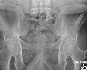
Figure 1: Bilateral sacroiliac joint posteroanterior radiograph obtained at the time of the current presentation.
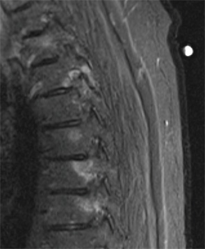
Figure 2: Sagittal STIR image from a thoracic spine MR, obtained two years prior to the current presentation. STIR sequences are fat-suppressed, water-sensitive images, useful for detecting bone marrow and soft tissue edema.
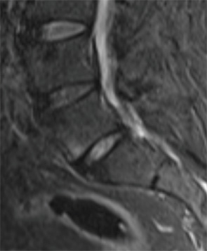
Figure 3: Sagittal STIR image from a lumbar spine MR, obtained two years prior to the current presentation.
