A 60-year-old Black woman with a history of stage 3 chronic kidney disease, type 2 diabetes and hypertension presented with a 12-month history of asymmetric polyarthritis of the wrists, metacarpophalangeal (MCP), proximal interphalangeal (PIP), metatarsophalangeal (MTP) and knee joints.
The review of systems was unremarkable. She denied oral ulcers, rashes, alopecia, or a history of pleural or pericardial effusions. She denied a history of psoriasis, dactylitis, inflammatory back pain, uveitis, abdominal pain, melena or hematochezia. There was no history of podagra, tophi or acute monoarthritis.
Laboratory studies were notable only for a positive anti-nuclear antibody at a titer of 1:80 with a homogenous pattern, and an elevated serum urate level of 9.5 mg/dL. She had chronic creatinine elevation, normocytic anemia and proteinuria attributed to diabetic nephropathy. Extractable nuclear antigens, complement levels, rheumatoid factor and anti-cyclic citrullinated peptide were negative.
In October 2018, treatment with adalimumab and prednisone was initiated with improvement of synovitis, but the patient’s disease flared upon tapering of the glucocorticoids. In July 2019, she presented to the clinic in tears, with acute monoarthritis of the right knee. She was unable to bear weight.
Arthrocentesis revealed inflammatory synovial fluid with 40,000 white blood cells/µL (neutrophil predominant). Gram stain, bacterial cultures and crystals were negative. She responded positively to glucocorticoids. Over the next year, her disease remained active in the hands, feet and knees, with minimal response to etanercept, tofacitinib or tocilizumab.
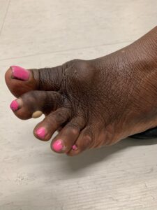
In October 2020, the patient returned to the clinic with hard, sub-centimeter, white lesions on her finger pads. A warm effusion of her left first MTP joint and a white blister on her left second toe were also noted on examination (see Figure 1). There was no sclerodactyly, Raynaud’s phenomenon or muscle weakness. Nailfold capillaries, muscle strength and creatinine kinase levels were normal.
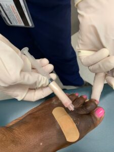
Aspiration of the first MTP joint and blister yielded 5 cc of chalky white fluid (see Figure 2). Polarized light microscopy revealed negatively birefringent crystals consistent with monosodium urate. Her serum urate level was 9.7 mg/dL. Repeat radiographs of both hands and feet showed interval erosive changes in both midfeet, consistent with gouty arthropathy.

FIGURE 2: Milk of urate aspirated from the first MTP joint and milk of urate bulla. (Click to enlarge.)
Allopurinol was initiated and titrated to a goal serum urate of less than 6.0 mg/dL, per the 2020 ACR Guideline for the Management of Gout.1 Five milligrams of prednisone by mouth daily was continued as flare prophylaxis.


