BALTIMORE—Although first described by the English physician Sir Jonathan Hutchinson in the 1870s, sarcoidosis remains a prevalent—and insidious—disease in the modern world.
At the 20th Annual Advances in the Diagnosis and Treatment of the Rheumatic Diseases Symposium at Johns Hopkins School of Medicine, Baltimore, Michelle Sharp, MD, MHS, assistant professor of medicine and co-director of the Johns Hopkins Sarcoidosis Program, provided a thorough lecture titled, Updates in Sarcoidosis.
Considerations for Diagnosis
First, Dr. Sharp noted three elements pertinent to correctly diagnosing sarcoidosis:
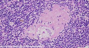
- The identification of non-caseating granulomatous inflammation on biopsy of an affected tissue;
- A clinical presentation compatible with the disease; and
- The exclusion of alternative etiologies for the patient’s signs/symptoms of disease.
Dr. Sharp stressed to the audience that the mere presence of non-caseating granulomas on a biopsy does not—in and of itself—make the diagnosis of sarcoidosis. Many entities may result in this histopathological finding, including lymphoma, aspergillosis, tuberculous and nontuberculous mycobacterial infection, histoplasmosis and vasculitis—to name just a few. Thus, it’s prudent to always think about and rule out infections and forms of malignancy while entertaining the diagnosis of sarcoidosis.
The typical onset of disease occurs between the ages of 20 and 40 years old, with a second peak occurring after age 60. (Note: This later onset is more common in women than men.) Although globally the incidence and prevalence of sarcoidosis is highest in Scandinavian and other Northern European countries, a three- to fourfold higher risk of disease exists for individuals who are Black vs. those who are white in the U.S.1 Being aware of these statistics is important, but Dr. Sharp advises physicians to avoid excluding the possibility of sarcoidosis in certain patients purely based on age, gender or race because any individual can be afflicted.
The immunologic hallmarks of sarcoidosis include: oligoclonal expansion of αβ+CD4+T cells, polarized Th1 immunity, regulatory T cell dysfunction and Th17/Th17.1 immunity. It has been hypothesized that microbes, including mycobacterium, may trigger this immunologic cascade; although it’s also thought that certain organic and inorganic agents may be implicated in pathogenesis of disease. This latter point has been emphasized by the observation of several studies that excess incident sarcoidosis occurred among the rescue and recovery workers exposed to debris following the World Trade Center attacks of Sept. 11, 2001.2,3
Dr. Sharp noted that sarcoidosis does not so much flare, but rather produces an undulating course of inflammation that may become clinically apparent when symptoms rise above the threshold noticeable to patients and physicians. If a patient with sarcoidosis is to go into remission, the vast majority do so within the first two or three years of their disease.
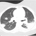
Many patients come to clinical attention based on imaging of the chest, which may demonstrate relatively symmetric and diffuse involvement of mediastinal and hilar lymph nodes, as well as thickened bronchovascular bundles. Unilateral pulmonary masses are rare, as are pleural effusions. When bronchoscopy is indicated, bronchoalveolar lavage (BAL) in patients with sarcoidosis would be expected to be predominantly lymphocytic.
Dr. Sharp pointed out that, in her clinical practice, she almost never orders a serum angiotensin-converting enzyme (ACE) level because the test is neither sensitive nor specific.
In the Organs
Sarcoidosis can affect nearly any organ in the body, and its presentations are often myriad. Cranial neuropathies can occur with involvement of the central nervous system, and Dr. Sharp has seen patients come to attention with the onset of acute hearing loss or dizziness.
Patients may also present with symptoms that mimic those of meningitis, encephalitis or transverse myelitis. If magnetic resonance imaging (MRI) of the brain is indicated to evaluate for evidence of leptomeningeal enhancement or other features of neurosarcoidosis, such imaging must be conducted with contrast to not miss these findings.
With regards to ocular involvement, uveitis is the most common finding. Cardiac involvement may include evidence of conduction delay, atrioventricular nodal block, bundle-branch block or fragmented QRS complexes.4 Cutaneous involvement of sarcoidosis can include classic findings, such as erythema nodosum or lupus pernio, as well as a wide range of skin lesions that have been described in association with the disease.
Patient Evaluation
Often, Dr. Sharp pursues all of the following tests when evaluating patients with sarcoidosis: computed tomography (CT) imaging of the chest, pulmonary function testing with spirometry and DLCO measurement at a minimum, electrocardiogram (ECG), complete blood count with differential, complete metabolic panel, measurement of calcium, 25-vitamin D, 1,25-vitamin D and referral to ophthalmology.
For assessing myocardial involvement, Dr. Sharp finds cardiac positron emission tomography (cardiac PET) more helpful than cardiac MRI because the latter cannot as easily distinguish between active disease and scarring. Even when directed at the heart, PET may pick up subclinical inflammation in other organs, such as the lungs or the liver.
Dr. Sharp explained that, although we typically think of patients with sarcoidosis as having restrictive or fibrotic lung disease, many other phenotypes are possible and should be screened for carefully. In a study of more than 600 patients with sarcoidosis, Dr. Sharp and her colleagues found that more than 50% had abnormal pulmonary function testing and that this finding included 22% with evidence of obstructive lung disease, 16% with an isolated reduction in DLCO and 15% with combined obstructive-restrictive lung disease.5
Treatment
No medications have been specifically approved for sarcoidosis by the U.S. Food & Drug Administration. However, corticosteroids remain the first-line treatment, with the dose and duration depending on the nature and severity of organ involvement.
Dr. Sharp explained that, for pulmonary involvement, 7.5–15 mg per day of prednisone may be sufficient and changes to therapy should be made slowly over time. Steroid-sparing agents include methotrexate, azathioprine, mycophenolate mofetil, tumor necrosis factor inhibitors and tocilizumab.
Although the field of sarcoidosis still has unmet needs, including the expansion of disease-targeted therapies, hearing from a “sarcoidologist”, such as Dr. Sharp, was helpful for the audience of rheumatologists. This old disease remains highly prevalent and relevant today, and talks like the one given by Dr. Sharp serve to keep the disease in the spotlight and ensure that the diagnosis is considered in all applicable situations.
Jason Liebowitz, MD, is an assistant professor of medicine in the Division of Rheumatology at Columbia University Vagelos College of Physicians and Surgeons, New York.
References
- Cozier YC, Berman JS, Palmer JR, et al. Sarcoidosis in Black women in the United States: Data from the Black Women’s Health Study. Chest. 2011 Jan;139(1):144–150.
- Crowley LE, Herbert R, Moline JM, et al. ‘Sarcoid like’ granulomatous pulmonary disease in World Trade Center disaster responders. Am J Ind Med. 2011 Mar;54(3):175–184.
- Izbicki G, Chavko R, Banauch GI, et al. World Trade Center ‘sarcoid-like’ granulomatous pulmonary disease in New York City Fire Department rescue workers. Chest. 2007 May;131(5):1414–1423.
- Cheng RK, Kittleson MM, Beavers CJ, et al. Diagnosis and management of cardiac sarcoidosis: A scientific statement from the American Heart Association. Circulation. 2024 May 21;149(21):e1197–e1216.
- Sharp M, Psoter KJ, Balasubramanian A, et al. Heterogeneity of lung function phenotypes in sarcoidosis: Role of race and sex differences. Ann Am Thorac Soc. 2023 Jan;20(1):30–37.