Findings/Diagnosis
The AP radiograph of the left hip (see Figure 1) shows periarticular, well-defined erosions of the left hip (white arrow) without joint space narrowing or osteophytes. There is no fracture. There are surgical clips and a calcified mass in the right hemipelvis (black ellipsis), representing a failed renal transplant.
The coronal STIR image of the pelvis demonstrates hypointense intra-articular synovial masses in the right and left hips (see Figure 2, white arrows). There are multiple synovial pressure erosions in the left acetabulum and femur (see Figure 3, white arrows); the articular cartilage otherwise appears preserved.
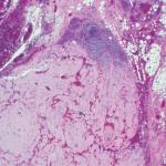
Based on these imaging findings and the history of chronic hemodialysis, the differential diagnosis included gout and amyloidosis. Given the polyarticular nature, pigmented villonodular synovitis was considered less likely. An ultrasound-guided synovial biopsy of the left hip was performed. Gram stain, culture and crystal analysis were negative. Surgical pathology revealed synovial tissue with nodular masses of extracellular, amorphous, eosinophilic material that was Congo-red positive, consistent with amyloid deposition. The patient had an elevated serum beta-2-microglobulin (25.8 mg/L; normal range 0.7–3.4 mg/L), further supporting the diagnosis.
Amyloid arthropathy is a known complication of long-term hemodialysis, characterized by the intraosseous, intra-articular and periarticular deposition of a unique form of amyloid composed of beta-2-microglobulin. This amyloid deposition in renal failure patients has a propensity for the musculoskeletal system, and the prevalence increases with the duration of dialysis therapy. The articular lesions are characterized by soft tissue masses, well-defined periarticular erosions and relative preservation of the joint space.
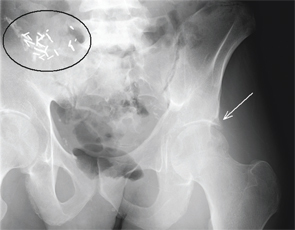
Figure 1.
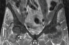
Figure 2.
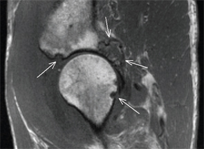
Figure 3.
Jennifer L. Demertzis, MD, is a musculoskeletal radiologist at the Mallinckrodt Institute of Radiology at Washington University School of Medicine in St. Louis, Mo. She is excited to collaborate on this new feature in the journal and looks forward to seeing future cases contributed by readers.
