SAN DIEGO—Rheumatologists are learning more about what autoantibodies, along with genetic and environmental factors, may predict the development of rheumatoid arthritis (RA) and systemic lupus erythematosus (SLE) years before symptoms appear. Speaking at the Clinical Research Conference at the 2013 ACR/ARHP Annual Meeting, held October 26–30, two researchers clarified recent findings in these areas that may one day enable rheumatologists to treat inflammation in a preclinical disease phase. [Editor’s Note: This session was recorded and is available via ACR SessionSelect at www.rheumatology.org.]
Paraphrasing a famous line from Shakespeare, Solbritt Rantapää-Dahlqvist, MD, professor of public health and clinical medicine at Umeå University, Umeå, Sweden, noted that in RA, “to be or not to be antibody positive, that is the question!” She and her colleagues looked at how the development of 10 critical antibodies to cyclic citrullinated peptides (CCP) might predict RA onset as many as seven years before clinical symptoms appear and what changes occurred in future RA patients’ immune responses up to a few weeks before disease onset. Their research was published in Arthritis & Rheumatism in April 2013.1
Telltale Autoantibodies
Using samples from the Medical Biobank of Northern Sweden, Dr. Rantapää-Dahlqvist and her colleagues analyzed the blood of 409 individuals, 386 of whom donated 717 samples before the onset of RA symptoms. For this group, there was a median of 7.4 years before RA symptom onset. In addition, blood biomarker data on 1,305 control subjects were analyzed. Antibodies to 10 citrullinated peptides were examined, but antibodies to three particular peptides stood out as highly predictive of RA: CEP-1, Fibß36-52, and filaggrin. In the samples studied, antibodies to these three peptides increased gradually over time, reaching their highest levels prior to RA disease onset. Fewer than 3.35 years before RA symptoms began, the odds ratio of a patient having both CEP-1 and Fibß36-52 was 40.4, compared to having antibodies to either peptide alone.
“We believe these antibodies might be important for the pathogenesis of the disease development” in RA, said Dr. Rantapää-Dahlqvist. In addition, between 3.4 and 0.2 years before RA starts, only 33.3% of patients showed no evidence of these antibodies in their blood, she said. “This would be a sign of epitope spreading, we think.”
The frequency of certain genetic isotopes that RA patients’ first-degree relatives have also may help rheumatologists predict disease before it strikes, Dr. Rantapää-Dahlqvist added. In another study, the Umeå researchers analyzed data from 51 families that included 163 people with RA and 157 first-degree relatives who did not have the disease. They found that both RA patients and their first-degree relatives had a high frequency of all anti-CCP and rheumatoid factors isotopes compared to unrelated, healthy individuals. In people with RA, the immunoglobulin (Ig) G isotype was most prevalent, but in their relatives, there was a high frequency of the IgA and IgM isotopes. The relatives’ blood rarely showed both anti-citrullinated peptide antibodies and rheumatoid factor, however.
“Despite the fact that these individuals did not fulfill the criteria of RA, they had ongoing processes that could lead to disease development,” Dr. Rantapää-Dahlqvist said. Individuals who did develop RA had a higher frequency of the human leucocyte antigen–shared epitope (HLA-SE) than their healthy relatives, and those who both smoked and had HLA-SE were significantly more likely to test positive for anti-CCP and rheumatoid factor.
Recent findings also show that, within two to four months of RA onset, patients will show a decrease in galactose levels as well as total IgG levels, Dr. Rantapää-Dahlqvist said. Another key autoantibody predicting RA onset may be anti–PAD-4; about 18.1% of patients test positive for it around five years prior to diagnosis, she added. Future research must clarify the role of all of these autoantibodies in the transition from autoimmunity to developing RA, she concluded. She believes that more data on first-degree relatives’ genetic makeup and smoking habits may provide key clues.
Epstein-Barr and Lupus
Lupus is another autoimmune disease whose preclinical clues are now being identified. Lupus is a “smorgasbord of clinical manifestations,” first written about by the ACR in 1972 to widespread ridicule, said John Harley, MD, PhD, director of rheumatology at Cincinnati Children’s Hospital. Later, identification of key genetic markers in SLE patients validated the findings. “The symptoms are unified by autoantibodies,” about 50 autoantibodies that SLE patients have in abundance, he said. “People with lupus come to the table with their own complement of risk or genetic predisposition, and then the environment comes into play.”
To identify who is at high risk of developing lupus before symptoms occur, researchers looked at the lupus patients and controls drawn from a U.S. Department of Defense repository that now contains over 40,000,000 serum samples and found key autoantibodies in patients who developed SLE five years later, Dr. Harley said. These patients developed autoantibodies and then had an asymptomatic period followed by typical lupus onset. Rheumatoid factor was associated with patients who developed arthritis, and anti-C1q and anti-dsDNA were associated with later renal disease, he noted. Another interesting finding was that about half of the African-American males who developed SLE had renal disease at the time of diagnosis and an increase in anti-dsDNA titer prior to disease onset. “The evidence is powerful that certain autoantibodies are needed for a particular disease expression,” Dr. Harley said.
In SLE, there is a reinforcing circle of autoimmune destruction, Dr. Harley said. Patients wind up with a life-threatening illness before treatments change its trajectory. Even a course of corticosteroids may alter the patient’s experience, he said. “Maybe if we could go back to the beginning and see what is happening, to the moment where they develop their first autoantibody, then we would understand the disease in a way that would empower us to be much more effective with our therapies.” Studies show that important preclinical markers of SLE that are the first to appear include anti–60kD-Ro, anti-dsDNA, and anti–nRNP-A. Over time, other autoantibody specificities accumulate in these patients, Dr. Harley added.
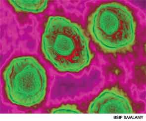
In two SLE autoantibody systems, the earliest autoantibody that appears to form also binds a protein from Epstein-Barr virus, a possible clue to the origin of autoimmunity in SLE. Studying the epidemiology of the Epstein-Barr virus may provide connections of this virus to SLE, Dr. Harley explained. However, 90% of adults carry Epstein-Barr, and most are infected between ages 12 and 16. So, Dr. Harley and his colleagues looked into seropositivity against Epstein-Barr in children.
Pediatric SLE patients mounted a more frequent and apparently different immune response to the Epstein-Barr nuclear antigen 1 (EBNA-1) antibody compared to control subjects, he said. SLE patients may, early on, develop autoantibodies directed against EBNA-1. In addition, there is a 15- to 40-fold increased Epstein-Barr virus load in patients with SLE, and an increased expression of Epstein-Barr–specific cells, he said. “The critical moment might be when the hetero-immune antibodies to EBNA-1 become autoantibodies binding to the SLE-specific antigens,” Dr. Harley noted. “It probably doesn’t take that much to make the transition to autoimmunity.”
Infection with the Epstein-Barr virus may be one of the key environmental risk factors for developing the autoimmunity of SLE, along with certain genes, sex, and ancestry. Vitamin D deficiency may also play a role, he noted. In conclusion, Dr. Harley recommended a clinical trial of hydroxychloroquine plus aspirin in preclinical SLE patients to determine whether this therapy would prevent SLE before the disease develops.
Susan Bernstein is a writer based in Atlanta.


