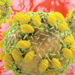
HBV, a de-emerging pathogen, is more complicated for patients with RA because of the threat of reactivation. Reactivation is potentially fatal but preventable, and it can occur after immunosuppression is either decreased or eliminated. This reactivation syndrome can occur in patients on low doses of methotrexate and prednisone, and it is increasingly reported in patients on biologic therapies. In the setting of HB surface antigen (HBsAg) positivity, treatment with a tumor necrosis factor inhibitor can lead to reactivation in as many as 40% of patients, Dr. Calabrese said.
All patients with a past history of HBV are at risk of reactivation, regardless of the presence or absence of serologic markers. Those at highest risk have chronic HBV infection, are HBsAg positive, and have high HBV DNA. At a bit lower risk are patients HBsAg positive and with either low or nondectable HBV DNA at baseline, he said. “My recommendation is that all patients commencing immunosuppressants and immunomodulatory therapies should be screened.”2
Diagnosing CNS Vasculitis
In his talk on CNS vasculitis, Dr. Calabrese noted that it is a treatable disease that can be diagnosed by presence of the following:
- A neurological sign or symptom (headache, stroke, seizure) that remains unexplained after a vigorous diagnostic workup;
- Some evidence of vascular disease in the brain, demonstrated by biopsy or angiography; and
- Exclusion of conditions capable of producing secondary arteritis or mimicking the angiographic features of arteritis.
Correct diagnosis is based on seven “rules of thumb,” he said. The first is that there is no clinical finding or set of findings of sufficient pretest probability to secure a diagnosis of primary angiitis central nervous system (PACNS) vasculitis.3 However, some signs can be definitive and, if present, should be elevated in a differential diagnosis: elevations of cells and proteins and normal glucose for three to six weeks after chronic infection and malignancy; recurrent focal deficits; constellation of both focal signs; and diffuse cortical impairment/neurological dysfunction.
Other tips for diagnosis:
- A lumbar puncture is the key laboratory test and should never be omitted unless there is some absolute neurosurgical contraindication.
- Normal magnetic resonance imaging (MRI) has a very high negative predictive value. “A combination of a normal MRI and normal lumbar puncture virtually rules out this diagnosis 100% of the time,” Dr. Calabrese said.
- No angiographic picture in the brain is 100% specific to the diagnosis of CNS vasculitis.
- Biopsy is an imperfect diagnostic tool, with a sensitivity of 75% or 80%, but it can rule out other conditions. “A nondiagnostic biopsy that has shown no evidence of infection or malignancy often is enough for me to be confident about investing in highly immunosuppressant therapy,” Dr. Calabrese said.
- Rule out infection and malignancy, and screen for viral infections, including HIV, HCV, and others.
- If the patient does not respond or progresses on cyclophosphamide and glucocorticoids, suspect an alternative diagnosis rather than refractory disease.
“Ask yourself at the end of your next consult whether any of these red flags are present,” Dr. Calabrese advised. “Was the diagnosis made in the absence of a lumbar puncture? Is the diagnosis made on angiogram with a normal lumbar puncture? And if the patient is not responding to aggressive therapy, reconsider the diagnosis.”

