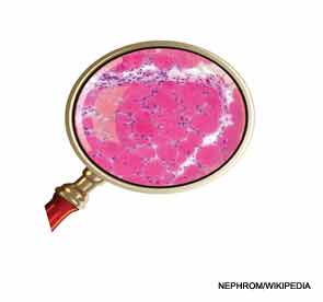
Tips for diagnosis and treatment of myositis and metabolic myopathies were highlighted in two presentations at the ACR 2013 State-of-the-Art Clinical Symposium, held April 20–21 in Chicago, with speakers underlining the need to recognize the clinical presentations of diseases that can mimic other conditions.
Paul H. Plotz, MD, scientist emeritus at the Arthritis and Rheumatism Branch of the National Institute of Arthritis and Musculoskeletal and Skin Diseases in Bethesda, Md., reminded attendees that many diseases can appear to be myositis. Mistaken diagnoses can result in antiinflammatory therapy for patients who will not benefit and may even suffer from the treatment, he said.
The first step in diagnosis of myositis is determining the location of the illness, whether it is actually inside or outside the muscle, and whether it is inflammatory, structural, or metabolic. The inflammatory myopathies include polymyositis, dermatomyositis, and inclusion body myositis (IBM).
Several clinical presentations should lead to a suspicion of the disease: Gottron’s or heliotrope rashes, a gradual onset of weeks to months, symmetry (except in the case of IBM), proximal limb and truncal involvement, Raynaud’s, arthritis, and idiopathic pulmonary fibrosis. Some myositic patients have musculoskeletal pain, but that is usually not the dominant feature of their illness, Dr. Plotz said. Most significantly, patients with myositis have elevated creatine kinase (CK), increasing weakness, and muscle edema that may be identified by magnetic resonance imaging (MRI). The MRI, however, will not identify the histology of the disease.
“Always biopsy a patient you suspect of having myositis. You are looking for lymphocytic information. Spend a moment and look at the biopsy yourself and convince yourself that there is truly inflammation and that there are some hints about whether it’s dermatomyositis or a polymyositis,” Dr. Plotz said, adding that dermatomyositis is a diagnosis made on examination of the skin for the characteristic rashes.
Several presentations should lead away from a diagnosis of myositis: a family history of a similar illness; weakness related to exercise, fasting, or eating; discrete episodes of reversible weakness; sensory, reflex, or other neurological signs; cranial nerve involvement; fasciculations; severe muscle cramping; myasthenia or myotonia; atrophy early in the disease or hypertrophy at any time; and CK that is normal or extremely high, he said.
Screening for Cancer
Patients with myositis, particularly those with dermatomyositis and polymyositis, develop cancer more frequently than the general population, with lung, ovarian, cervical, colorectal, gastric, breast, bladder, and pancreatic cancer topping the list of the most frequent.1 A middle-aged woman with dermatomyositis has a 2- to 4-fold risk of cancer within a year of her diagnosis of dermatomyositis and 1.5- to 2-fold risk if she has polymyositis. Patients most at risk of developing cancer are older than age 45. The cancer risk falls back to baseline risk within about five years of diagnosis of myositis, he said.


