 Rheumatic Diseases of Childhood: Juvenile Dermatomyositis with Calcinosis Cutis
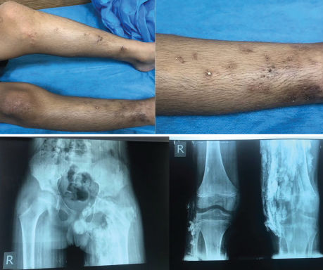
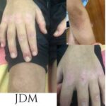
These images depict a 14-year-old boy with a two-year history of proximal muscle weakness affecting both upper and lower limbs, and a skin rash affecting his face. He was diagnosed with juvenile dermatomyositis and developed calcinosis over both legs with skin infection and ulceration. Plain X-ray of both his pelvis and femurs showed reticular pattern calcinosis. He had a poor response to a infliximab.
Hala Fadhil Hasan, MBChB, is a rheumatology fellow at Baghdad Teaching Hospital, Iraq, and a lecturer and faculty member of Alkindy Medical College, University of Baghdad.
About the Contest
The Rheumatology Image Library is a highly accessed teaching resource. However, images showing manifestations of rheumatic disease on skin of color are under-represented, creating a significant educational gap. The 2021 Image Competition, held in conjunction with ACR Convergence 2021, encouraged the global rheumatology community to submit images that will help healthcare providers identify rheumatic disease manifestations in skin of color. Here, we depict the image featured from the Middle East & North Africa. In coming issues, we’ll publish the winning images from other regions. The images judged Best Overall and People’s Choice, and those featured from East Asia & the Pacific Region, as well as South Asia, appeared in previous issues.

