Periungual Erythema & Its Translation on Nailfold Videocapillaroscopy in a Patient with Very Early Systemic Sclerosis
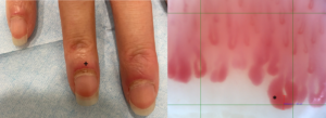
A 66-year-old woman presented with Raynaud’s phenomenon and periungual erythema. HEp-2 immunofluorescence assay was positive for antinuclear antibodies, showing a centromere pattern. The presence of anti-centromere antibodies was confirmed by chemiluminescent immunoassay. The patient was diagnosed with very early systemic sclerosis. The image on the left shows periungual erythema (+) visible in the third, fourth and fifth fingers of the right hand. The image on the right exhibits its translation as giant capillaries (*) on nailfold videocapillaroscopy of the fourth finger of the right hand, corresponding to an active scleroderma pattern.
Joana Martins Martinho, MD, is a rheumatologist resident in the Rheumatology Department at the Centro Hospitalar Universitário Lisboa Norte, Lisbon, Portugal.
About the Contest
The Rheumatology Image Library is a highly accessed teaching resource. The 2022 Image Competition sought images representing a diverse range of patients who show either characteristic or unusual manifestations of rheumatic disease, including systemic sclerosis, localized scleroderma and scleroderma mimics. Look for the Best Overall Image on our website and other regional winners in future issues or in the Rheumatology Image Library.


