Tumoral Calcinosis in Systemic Sclerosis
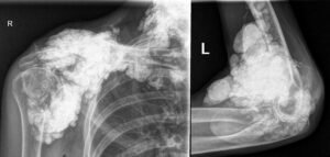
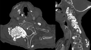
Plain radiograph and computed tomography images depict calcinosis in the region of the right shoulder, left elbow and cervical spine of a 71-year-old woman with a 20-year history of systemic sclerosis. Manifestations include high-titer anti-Scl-70 antibody, diffuse skin involvement, Raynaud’s syndrome, acro-osteolysis, gastroesophageal reflux disease, interstitial lung disease and complete heart block.
Recently, extensive tumoral calcinosis has been the most challenging manifestation of her systemic sclerosis due to chronic pain and impairment of functional activities.
Jonathan T. Cheah, MBBS, is an assistant professor of medicine in the Division of Rheumatology, UMass Chan Medical School, Worcester, Mass.
About the Contest
The Rheumatology Image Library is a highly accessed teaching resource. The 2022 Image Competition sought images representing a diverse range of patients who show either characteristic or unusual manifestations of rheumatic disease, including systemic sclerosis, localized scleroderma and scleroderma mimics. Look for the Best Overall Image on our website and other regional winners in future issues or in the Rheumatology Image Library.


