Diffuse Cutaneous Scleroderma with Interstitial Lung Disease: Radiological Findings
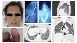
A 44-year-old woman with cutaneous manifestations, tightening of her face with a reduced oral aperture and a healing digital tip ulcer (Figures 1 and 2) has had a known case of diffuse scleroderma since 2008. The patient presented with complaints of shortness of breath along with dysphagia. A barium swallow showed dilated distal thoracic esophagus without mural mass (Figure 3). A high-resolution computed tomography (HRCT) chest scan (Figures 4, 5 and 6) showed areas of inter- and intra-lobular septal thickening with tractional bronchiectasis, honeycomb cysts and groundglassing in apico-basilar gradient suggestive of interstitial lung disease (UIP pattern). A dilated esophagus was also shown, suggestive of motility disorder related to scleroderma.
Shung Ming Chiu, MBBS, is a medical officer in rheumatology, a member of the Spondylitis Association of America and its first International Support Group Leader. He has previously written for The Rheumatologist about his experience completing medical training with a diagnosis of spondyloarthritis.
About the Contest
The Rheumatology Image Library is a highly accessed teaching resource. The 2022 Image Competition sought images representing a diverse range of patients who show either characteristic or unusual manifestations of rheumatic disease, including systemic sclerosis, localized scleroderma and scleroderma mimics. Look for the Best Overall Image on our website and other regional winners in future issues or in the Rheumatology Image Library.


