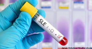
 Autoantibodies that react with long interspersed nuclear elements (LINE-1) p40 protein may be common in severe and active systemic lupus erythematosus (SLE) patients, according to research from Victoria Carter, MS, and colleagues.1
LINE-1, class I transposable elements in DNA, make up 17% of the human genome. Although most LINE-1 copies are mutated and truncated in the human genome, approximately 180 appear to be intact, with a few that continue to retrotranspose and occasionally disrupt genes or regulatory regions by novel insertions, according to the researchers. Because of this response, defense mechanisms have evolved against retro-elements and retroviruses. Some researchers theorize that many human diseases, including cancer and autoimmune diseases, are connected with LINE-1 biology, and several theories link LINE-1 to the development of SLE and its flares.
To help test this hypothesis, researchers used highly purified p40 proteins to quantitate immunoglobulin G autoantibodies found in the serum of 172 SLE patients, as well as disease controls (20 patients with systemic sclerosis) and age-matched healthy subjects (78 patients). The first cohort for the study included 10 patients with SLE. In a second SLE cohort, patients were in remission (n=83) or experiencing flare (n=79). The initial group of 10 patients came from the University of Washington Medical Center, Seattle, and the remaining patients were from Skåne University Hospital, Lund, Sweden.
In the majority of patients, as well as in many healthy controls, antibodies reactive with p40 proteins were detected. The results were higher in SLE patients, but not those with systemic sclerosis, when compared with healthy controls. Anti-p40 reactivity was higher during a flare among patients in remission (P=0.03). Anti-p40 reactivity also correlated with several other factors, including Systemic Lupus Erythematosus Disease Activity Index (SLEDAI) (P=0.0002), type I interferon score (P=006), complement C3 decrease (P=0.0001), anti-DNA antibodies (P<0.0001), anti-C1q antibodies (P=0.004) and a current or past history of nephritis (P=0.02). The antibodies were correlated inversely with age. In a study of SLE patient sera, the researchers also found a reaction with p40-associated proteins.
Within the SLE flare population, patients with high titers had higher SLEDAI scores than patients who were below the cutoff of the 90th percentile or more among healthy controls. Current anti-dsDNA antibodies and complement consumption also were associated with anti-p40 antibody levels. “Collectively, these data indicate that higher anti-p40 levels tend to accompany active disease,” the researchers write.
One finding that surprised researchers is a statistically higher inverse correlation of anti-p40 reactivity with SLE patient age (P<0.0001), which may be partially explained by the higher SLEDAI of younger patients, they write. However, even when the full cohort of SLE patients and healthy controls was split into two groups with an age cutoff at 40 years, researchers found a more marked association of anti-p40 reactivity with SLE in the younger group than in the total cohort.
LINE-1 may be a key trigger of SLE, working along with Ro60, La & other RNA binding proteins, according to Tomas Mustelin, MD, PhD.
Future Implications
“Clearly, anti-p40 antibodies do not by themselves herald clinically relevant autoimmunity, but more likely represent an early phase of self-reactivity that may—or may not—progress towards SLE. … Our findings that nearly all SLE patients have autoantibodies against the LINE-1 p40 protein and that these antibodies are associated with disease activity, specific disease manifestations, low complement, other autoantibodies and type I interferons, taken together, suggest that LINE-1 biology is coupled in some way to SLE pathogenesis,” the researchers write.
However, this does not necessarily mean LINE-1 cause SLE. Instead, it may be targeted by the immune response as, what the researchers call, “an innocent bystander.”
“The physical interaction of p40 with well-known SLE autoantigens would be compatible with such a role, at least if one assumes that Ro and La are the intended antigens for the immune response,” the researchers write.
The researchers hypothesize that LINE-1 may be a key trigger of the disease, working along with Ro60, La and other RNA binding proteins, according to study co-author Tomas Mustelin, MD, PhD, professor of medicine, Herndon and Esther Maury Endowed Professor in Rheumatoid Arthritis, Division of Rheumatology, Department of Medicine, University of Washington, Seattle.
Depending on future research results, LINE-1 p40 autoantibodies could become part of the lab tests used to assess SLE patients or could be a biomarker in clinical trials of new targeted lupus therapies, Dr. Mustelin says.
The study authors are encouraged by the potential therapeutic implications of their hypothesis, with Dr. Mustelin adding that some of the reverse ranscriptase inhibitors used to treat HIV, which also block the reverse transcriptase activity of LINE-1 p145, would likely stop type I interferon production, potentially tempering SLE disease.
“Our goal is to directly test this possibility,” he says.
Other research must first take place, and one study the authors have underway is analyzing LINE-1 p40 autoantibodies in pediatric lupus patients and in those with other autoimmune diseases that share a type I interferon signature with SLE.
Chandra Mohan, MD, PhD, professor of biomedical engineering and medicine, Cullen College of Engineering, University of Houston, and a Lupus Foundation of America Medical-Scientific Advisory Council member, found the study results promising. “Although there has been accumulating evidence describing the importance of LINE-1 in SLE, this report brings a novel perspective to this field, with a focus on autoantibodies targeting LINE-1 elements,” Dr. Mohan says.
Future studies with independent patient cohorts are needed to validate the researchers’ observations, including a comparison of current diagnostic tests, with careful assessment of sensitivity and specificity, Dr. Mohan notes. Another approach would be longitudinal studies to examine whether the autoantibodies are definite early harbingers of SLE or disease flare.
Mary K. Crow, MD, chair, Department of Medicine, Benjamin M. Rosen Chair in Immunology and Inflammation Research, Hospital for Special Surgery, and chief, Division of Rheumatology, Joseph P. Routh Professor of Rheumatic Diseases in Medicine, Weill Cornell Medical College, New York, has previously reported on the connection between LINE-1 and the pathogenesis of SLE.2,3 Some of her previous work supports the potential role for LINE-1 in some patients with SLE and a link between LINE-1 and type 1 interferon production. However, she believes the connection is still speculative. In a letter to the editor regarding this study, she noted that LINE-1 p40 and other proteins implicated as autoantigens in systemic autoimmune disease should be priority research targets.4
Vanessa Caceres is a medical writer in Bradenton, Fla.
References
- Carter V, LaCava J, Taylor MS, et al. High prevalence and disease correlation of autoantibodies against p40 encoded by long interspersed nuclear elements in systemic lupus erythematosus. Arthritis Rheumatol. 2020 Jan;72(1):89–99.
- Crow MK. Long interspersed nuclear elements (LINE-1): Potential triggers of systemic autoimmune disease. Autoimmunity. 2010 Feb;43(1):7–16.
- Mavragani CP, Sagalovskiy I, Guo Q, et al. Long interspersed nuclear element-1 retroelements are expressing in patients with systemic autoimmune disease and induce type 1 interferon. Arthritis Rheumatol. 2016 Nov;68(11):2686–2696.
- Crow MK. Reactivity of IgG with the p40 protein encoded by the long interspersed nuclear element 1 retroelements: Comment on the article by Carter et al. Arthritis Rheumatol. 2020 Feb;72(2):374–376.

