I had a patient at the Veteran’s Administration (VA) hospital whom I frequently treated during the last two months of my rotation at that facility. While I need not share the specific details of his illness, the patient is perhaps the most important component of this musculoskeletal ultrasound (MSUS) story.
My experience with ultrasound (US) did not begin at the VA. I generally was familiar with this technique before starting my fellowship. Using US as the joints’ imaging modality emerged in American rheumatology over a decade ago. MSUS uses the concept of tissue echogenicity, which refers to the ability to reflect or transmit US waves against the surrounding tissues. The difference in tissue context—echogenicity—is visible on the US monitor as a change in contrast. Based on echogenicity, commonly referred to as gray scale, a structure can be defined as hyperechoic, seen as white on the screen, hypoechoic gray and anechoic black (see Figure 1, opposite). MSUS can augment recognition of active inflammation with the use of color Doppler where increased signal identifies better perfused, diseased tissue.
When I became aware that the VA had a US machine available for rheumatology use, I became interested in gaining some experience with the technique during my rotation there. My patient proved not only an enthusiastic model for scanning, but also an agent provocateur, stimulating my interest in polishing my skills with this technique.
Key to Abbreviations
- ACR’s RhMSUS: The American College of Rheumatology’s Musculoskeletal Ultrasound Certification in Rheumatology
- MSUS: musculoskeletal ultrasound
- PAN: polyarteritis nodosa
- UCLA: University of California in Los Angeles
- UTSW: Texas Southwestern University
- VA or VAMC: Veterans Affairs Medical Center
Patient Response to US Years ago my patient was diagnosed with polyarteritis nodosa (PAN) that doesn’t follow any pattern or respond to any treatment. His situation was one of those for which no trial helped guide me toward a choice of therapy, no expert opinion moved me forward, no state-of-the-art lecture provided me with an “aha moment.” His illness kept me awake at night.
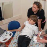
Ultrasound machines are expensive. Their luxury-car price tag explains why the one at the VA was garaged in the office of the chief of rheumatology, located on the fifth floor. Three mornings a week, I wrestled with the machine, transporting it down four floors to the exam room, in and out of elevators, zig-zagging between the waiting room seats, trying to avoid inflicting damage to any federal property. One day, my patient saw me rolling this cumbersome machine into the exam room and commented on “what a nice toy” it seemed to be. I explained how the US beam allowed me to see small fragments of tissue. His interest piqued, my patient immediately volunteered to be scanned. As a veteran and a beneficiary of the VA medical system, he saw himself as part owner of the machine and felt obliged to help me maneuver the machine.
