“We are struggling to find what could be the correct range where we think this could still be a normal background noise or a normal reference range, not to overdiagnose spondyloarthritis,” says Dr. Weber. “We think the present proposal of two lesions is too low.”
Researchers reviewed data from MRI scans of sacroiliac joints of 20 recreational runners (12 women, eight men) and a group of 22 professional male hockey players from the Danish Premier League. All were healthy athletes ranging in age from 18 to 40 years old.
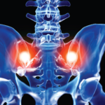
The study explored the frequency and anatomic distribution of sacroiliac joint lesions on MRI to determine the intersection between normal variation and disease. Researchers selected recreational and elite athlete groups to investigate extremes of axial skeletal strain—low intensity in the runners and maximum intensity in the professional ice hockey players, says Dr. Weber.
“We wanted to compare those two extremes and look at how many of these obviously healthy persons would be (given) a diagnosis of sacroiliitis with the current MRI definition. … So how many false positives would we create in persons with this high axial strain?” he asks.
The scans were conducted on the runners before and after a 6.2 km run and on the hockey players at the end of their sport season. Independent blinded readers analyzed the MRI data and recorded subjects who met Assessment of SpondyloArthritis International Society (ASAS) definition of active sacroiliitis. Mixed in with the reviewed data to serve as masking were seven MRI scans (with two paired images) of spondyloarthritis patients who were undergoing anti-tumor necrosis factor (anti-TNF) treatment.
Descriptive analysis included the mean frequency of sacroiliac joint quadrants exhibiting bone marrow edema and structural lesions and their distribution in eight anatomic sacroiliac joint regions that involved the upper and lower ilium and sacrum, subdivided into anterior and posterior slices.
Results

Dr. Deodhar
The results indicated that bone marrow edema was common among both cohorts of healthy athletes, showing up in the MRI scans of an average of three to four joint quadrants per subject, meeting the ASAS definition of active sacroiliitis in 30–41% of study participants, Dr. Weber says. The posterior lower ilium was the single most affected sacroiliac joint region.
Joint erosion was virtually absent in the scans of study participants, yet one in three of these healthy athletes met the most widely applied classification criteria for sacroiliitis based solely on bone marrow edema, says Dr. Weber. This supports the authors’ argument that the current threshold recommended by ASAS of at least two lesions is not enough and the bar to reach diagnosis of spondyloarthritis needs to be set higher.

