This case is a clear example of early SSc (limited SSc); even in the absence of other non-Raynaud’s symptoms of disease, the diagnosis of SSc was possible because of the appearance of the classical morphological markers for microvessel damage (early NVC scleroderma pattern) that can characterize the beginning of the disease and have been validated.4
The Role of Microvascular Damage Assessment in SSc
SSc is a rare progressive connective tissue disease characterized by vascular abnormalities/damage that is associated with diffuse fibrosis in the skin and progressive involvement of the internal organs. The origin of SSc appears multifactorial, although the patient’s gender has a strong impact. SSc is about eight times more common among women than men; the dramatic female predominance of the disease may reflect the role of estrogens in enhancing the immune responses.5 Most common in the third to fifth decades of life, SSc is rare in children. Although the basis for the age distribution is unknown, intriguing studies suggest a role of the aromatases in the periphery as a source of estrogen metabolites to enhance cell proliferation and fibrosis.6
The most common initial symptoms and signs of SSc are vascular, such as Raynaud’s phenomenon, which is often long lasting, as well as insidious swelling of the distal extremities (e.g., puffy fingers). These features are followed by gradual thickening of the skin of the face and fingers along with skin ulcers (see Figures 3 and 4). The diagnosis of SSc in its earlier stage is clinical and includes the presence of Raynaud’s phenomenon together with capillaroscopic markers of microvasculature damage (e.g., giant capillaries and related microhemorrhages); biomarkers, namely SSc-specific serum autoantibodies, can help confirm the diagnosis.
Does Microvascular Change Reflect Pathophysiology?
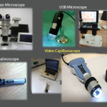
Peripheral microvascular damage in SSc is characterized by dynamic alterations in capillary structure that eventually can lead to a progressive decrease in capillary density. In the nailfolds, where the progression of the morphological changes can be detected by safe and noninvasive videocapillaroscopy (a microvideocamera, see Figure 5), capillary loops dilate homogenously (giant capillary) as they are damaged by the local immune/inflammatory reaction and gradually lose their red blood cell content (microhemorrhage); the loss of microvascular loops begins with their progressive collapse (see Figure 6). The reason that Raynaud’s phenomenon can produce an early NVC pattern relates to the effects of local ischemia from vasoconstriction; over time, this process may induce endothelial cell and tissue necrosis and damage (see Figure 7). The dynamic involvement of capillaries by the immunopathological process in SSc may be associated with worsening of the capillaroscopic pattern (from early to active pattern) that can be linked to overt disease symptoms (e.g., skin trophic lesions) (see Figure 8 and 9).

