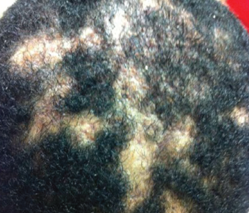
Figure 1. Dissecting cellulitis of the scalp. Note the areas of alopecia where subcutaneous abscesses are located.
Synovitis, acne, pustulosis, hyperostosis and osteitis (SAPHO) syndrome is a heterogeneous, inflammatory, musculoskeletal disease. The disease is an insidious, sterile osteitis with associated skin and synovial inflammation.1 Diagnosis can prove challenging, but a thorough clinical history, high clinical suspicion and imaging techniques can help clinch it.
The below case reveals a rare, fulminant presentation of SAPHO syndrome in which signature imaging findings helped us achieve diagnosis. It highlights an unusual combination of multilevel spinal involvement, scalp and extremity dissecting cellulitis, and osteitis with the signature bull’s head sign on scintigraphy. This unique presentation required excluding mimics, such as spondyloarthropathy, infection, malignancy and other immunodeficiencies.
The Case
A 28-year-old black man arrived at our rheumatology clinic in a wheelchair pushed by his mother. He appeared very stiff and uncomfortable and had been confined to the wheelchair for about eight weeks prior to his arrival.
He was in marked distress as he and his mother described his history. He developed acute onset of back and neck pain about 10 weeks prior to his visit. About six weeks prior to his presentation, he developed arthritis of the knees and left shoulder, as well as a scaly rash on his left lower leg and scalp. The rash on his scalp progressed to itching and oozing lesions. He completely lost his appetite, which was associated with a 30 lb. weight loss since the onset of symptoms. He denied any fever, but did describe subjective chills and constant fatigue. After the onset of arthritis, he became almost entirely confined to the wheelchair because the pain and stiffness became so severe that weight bearing was difficult. In fact, it was painful to sit upright in the wheelchair while his mother navigated him from place to place for his daily activities.
He had no significant past medical or surgical history and took no medications. He reported consuming large amounts of acetaminophen and ibuprofen daily to alleviate his severe pain and discomfort. He denied a family history of inflammatory joint disease, psoriasis or malignancy. He denied ever using illicit substances, drinking alcohol, using tobacco products or recent travel.
He was afebrile, but on examination appeared ill and in obvious discomfort. He was so stiff he resembled a statue sitting in the wheelchair. His scalp featured multiple, purulent, draining areas of fluctuance erythema and edema (see Figure 1). Palpation of his neck revealed bilateral, small, tender lymph nodes. The rash on his left lower leg was less purulent and more eczematous and scaly on an erythematous base.

