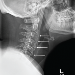
Next, multifocal areas of vertebral bone marrow edema consistent with osteitis appear on MRI (see Figures 2A and 2B). Vertebral lesions are classically described as corner lesions owing to the bone edema usually associated with erosion found at the anterior corners of the vertebral bodies (see Figure 2B). These lesions are difficult to differentiate from infectious and malignant bone lesions, so biopsy is usually required to evaluate the differential diagnoses.
Hyperostosis noted on plain X-ray can appear and is most commonly noted in the clavicle (although the radiographs of our patient revealed hyperostosis of the proximal humerus and distal acromion). Finally, in contrast to hyperostosis lesions, osteolytic lesions may be discovered as well.6,10
Our patient’s presentation was unique because it included all the typical imaging findings of SAPHO, and this constellation of radiographic findings was key in clinching the diagnosis.
Conclusion
SAPHO syndrome remains a poorly understood disease and is thought, at this time, to be rare. The literature describing SAPHO suggests it can present in a myriad of ways. Our case was unique because it occurred in a black man and featured fulminant onset, all the classic radiographic findings and a rarely associated dissecting cellulitis dermatosis.
Several treatment options are available for SAPHO. Our patient’s fulminant course was successfully treated with anti-TNF therapy. Physical therapy, topical steroids and antibiotics for skin lesions add to the therapeutic armamentarium.
Still, we need larger cohorts and collaborative studies to better define efficacy and outcomes in the management of SAPHO syndrome.
 Ross J. Thibodaux, MD, is a rheumatology fellow in the Division of Rheumatology at the Louisiana State University Health Science Center in New Orleans.
 Nirupa J. Patel, MD, is an associate professor in the Division of Rheumatology at the Louisiana State University Health Science Center in New Orleans.
References
- Firinu D, Garcia-Larsen V, Manconi PE, et al. SAPHO syndrome: Current developments and approaches to clinical treatment. Curr Rheumatol Rep. 2016 Jun;18(6):35.
- Rukavina I. SAPHO syndrome: A review. J Child Orthop 2015 Feb;9(1):19–27.
- Aljuhani F, Tournadre A, Tatar Z, et al. The SAPHO syndrome: A single-center study of 41 adult patients. J Rheumatol. 2015 Feb;42(2):329–334.
- Li C, Zuo Y, Wu N, et al. Synovitis, acne, pustulosis, hyperostosis and osteitis syndrome: A single centre study of a cohort of 164 patients. Rheumatol (Oxford) 2016 Jun;55(6):1023–1030.
- Colina M, Govoni M, Orzincolo C, et al. Clinical and radiologic evolution of synovitis, acne, pustulosis, hyperostosis, and osteitis syndrome: A single center study of a cohort of 71 subjects. Arthritis Rheum. 2009 Jun 15;61(6):813–821.
- Nguyen MT, Borchers A, Selmi C, et al. The SAPHO syndrome. Semin Arthritis Rheum. 2012 Dec;42(3):254–265.
- Benhamou CL, Chamot AM, Kahn MF. Synovitis-acne-pustulosis-hyperostosis-osteomyelitis syndrome (SAPHO). A new syndrome among the spondyloarthropathies? Clin Exp Rheumatol. 1988 Apr–Jun;6(2):109–112.
- Kahn MF, Khan MA. The SAPHO syndrome. Baillieres Clin Rheumatol. 1994 May;8(2):333–362.
- Zwaenepoel T, Vlam KD. SAPHO: Treatment options including bisphosphonates. Semin Arthritis Rheum. 2016 Oct;46(2):68–173.
- Depasquale R, Kumar N, Lalam RK, et al. SAPHO: What radiologists should know. Clin Radiol. 2012 Mar;67(3):195–206.

