The rest of his laboratory testing results were unremarkable: His HLA-B27 was negative, as were his anti-nuclear antibody (ANA) titers and rheumatoid factor levels. Other serologies, including antibodies against cyclic citrullinated peptide (CCP), SSA/Ro, SSB/La, nuclear ribonucleoprotein (RNP), Smith antigen and double-stranded DNA were negative. Additionally, C3 levels, C4 levels and ferritin levels were within normal limits, and HIV was non-reactive.
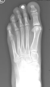
X-rays of both hands showed acro-osteolysis of the distal phalanges of all digits. X-rays of both feet showed soft tissue calcinosis and resorption of the distal phalanges (see Figures 2 and 3).
Electromyography (EMG) showed a partial radial mononeuropathy in the right upper extremity, distal to the spiral groove. Preservation of the distal motor amplitude suggested favorable prognosis for spontaneous recovery; however, there was a mild distal axonal sensory peripheral polyneuropathy. Of note, a 9–10 Hz tremor was observed in several muscles.
Discussion
When we saw this patient, we investigated rheumatic etiologies for acro-osteolysis, especially given the pains he was experiencing; however, our workup was nonrevealing. Given the patients severe osteoporosis, the suspicion of HCS was high. His pain, swelling and neuropathic symptoms were likely secondary to HCS.

FIGURE 3: Left foot X-ray shows calcinosis in the
second distal phalanx and resorption of distal phalanges. (Click to enlarge.)
Acro-osteolysis can occur in a variety of conditions, but rheumatologists are most likely to encounter acro-osteolysis in systemic sclerosis and psoriatic arthritis. It can also be found in rheumatoid arthritis (RA), granulomatosis with polyangiitis (GPA), systemic lupus erythematosus (SLE) and Sjögren’s disease.1 Acro-osteolysis can occur in up to 65% of patients with systemic sclerosis, depending on the definition and the sensitivity of the confirmatory imaging modality.2 The pathophysiology in scleroderma appears to be related to microangiopathy causing hypoxia and leading to bone resorption and remodeling.6
Regarding psoriatic arthritis (PsA), a case series found three of 47 patients had acro-osteolysis.5 Psoriatic arthritis involves erosive changes in the distal extremity joints and the chronic inflammation associated with PsA is thought to cause the bone destruction found in acro-osteolysis.3
In RA, GPA, SLE and Sjögren’s disease, acro-osteolysis has been described in case reports.
Non-rheumatic etiologies of acro-osteolysis include occupational exposure, bacterial infections, hyperparathyroidism, diabetes mellitus type 1, lysosomal disease and other genetic disorders, such as Gaucher’s disease.2,4
Diagnosis of HCS includes genomic sequencing. Current investigational treatments are osteoporotic medications, but it is unclear if these are helpful.1 Our patient was offered zoledronic acid, but he was lost to follow-up.


