Hajdu-Cheney syndrome (HCS) is a rare connective tissue disease, with fewer than 100 cases reported worldwide.1 Hallmark features include acro-osteolysis (i.e., resorption of the distal phalanges of the hands and feet), osteoporosis, facial dysmorphisms, and craniofacial and dental abnormalities. Patients often have short stature and can have neuroanatomical deformities causing intellectual disabilities. These patients can also have atrioventricular septal defects and polycystic kidneys. HCS is inherited in an autosomal-dominant pattern and is associated with mutations in the NOTCH-2 protein, although the exact mechanism is still under investigation.1 HCS is not only uncommon, but it can present in various ways, making it important to consider in differential diagnoses of several diseases.
We present the case of a young man with upper and lower extremity joint pain and acro-osteolysis presenting for rheumatic evaluation.
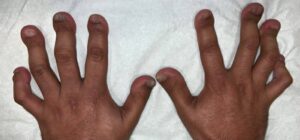
FIGURE 1: Both hands show boutonniere deformities in the third and fourth fingers, with onychorrhexis or longitudinal ridging. (Click to enlarge.)
Case Presentation
Our patient is a 29-year-old man with a past medical history of acro-osteolysis and osteoporosis who presented to our rheumatology clinic with diffuse joint pain. His pain is usually in his right arm in the bicep area and radiates to the forearm. He has associated neuropathy symptoms in both hands. Although his symptoms usually manifest in his right upper extremity, he has had similar symptoms in his left upper and both lower extremities as well. These symptoms occur every few years, persist for a few months and then remit without treatment. He does not have alleviating or aggravating factors and there were no known triggers.
On physical exam, the patient did not have any active synovitis, skin changes or strength deficits. He did have shortened, clubbed digits and flexion deformities of the right fourth and fifth proximal interphalangeal joints. The patient also had a boutonniere deformity in both the third and fourth digits (see Figure 1).
Laboratory studies showed a normal urinalysis and his hemoglobin mildly elevated at 17.7 g/dL (reference range [RR]: 12–15 g/dL). A comprehensive metabolic panel was significant for mildly elevated aspartate transaminase at 43 u/L (RR: 13–39 u/L) and alanine transaminase at 69 u/L (RR: 7/52 u/L). He also had an elevated uric acid at 8.1 mg/dL (RR: 1.5–6.5 mg/dL) and positive anti-mitochondrial M2 antibody at 40.2 u.
The rest of his laboratory testing results were unremarkable: His HLA-B27 was negative, as were his anti-nuclear antibody (ANA) titers and rheumatoid factor levels. Other serologies, including antibodies against cyclic citrullinated peptide (CCP), SSA/Ro, SSB/La, nuclear ribonucleoprotein (RNP), Smith antigen and double-stranded DNA were negative. Additionally, C3 levels, C4 levels and ferritin levels were within normal limits, and HIV was non-reactive.
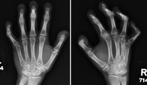
X-rays of both hands showed acro-osteolysis of the distal phalanges of all digits. X-rays of both feet showed soft tissue calcinosis and resorption of the distal phalanges (see Figures 2 and 3).
Electromyography (EMG) showed a partial radial mononeuropathy in the right upper extremity, distal to the spiral groove. Preservation of the distal motor amplitude suggested favorable prognosis for spontaneous recovery; however, there was a mild distal axonal sensory peripheral polyneuropathy. Of note, a 9–10 Hz tremor was observed in several muscles.
Discussion
When we saw this patient, we investigated rheumatic etiologies for acro-osteolysis, especially given the pains he was experiencing; however, our workup was nonrevealing. Given the patients severe osteoporosis, the suspicion of HCS was high. His pain, swelling and neuropathic symptoms were likely secondary to HCS.
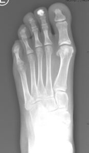
FIGURE 3: Left foot X-ray shows calcinosis in the
second distal phalanx and resorption of distal phalanges. (Click to enlarge.)
Acro-osteolysis can occur in a variety of conditions, but rheumatologists are most likely to encounter acro-osteolysis in systemic sclerosis and psoriatic arthritis. It can also be found in rheumatoid arthritis (RA), granulomatosis with polyangiitis (GPA), systemic lupus erythematosus (SLE) and Sjögren’s disease.1 Acro-osteolysis can occur in up to 65% of patients with systemic sclerosis, depending on the definition and the sensitivity of the confirmatory imaging modality.2 The pathophysiology in scleroderma appears to be related to microangiopathy causing hypoxia and leading to bone resorption and remodeling.6
Regarding psoriatic arthritis (PsA), a case series found three of 47 patients had acro-osteolysis.5 Psoriatic arthritis involves erosive changes in the distal extremity joints and the chronic inflammation associated with PsA is thought to cause the bone destruction found in acro-osteolysis.3
In RA, GPA, SLE and Sjögren’s disease, acro-osteolysis has been described in case reports.
Non-rheumatic etiologies of acro-osteolysis include occupational exposure, bacterial infections, hyperparathyroidism, diabetes mellitus type 1, lysosomal disease and other genetic disorders, such as Gaucher’s disease.2,4
Diagnosis of HCS includes genomic sequencing. Current investigational treatments are osteoporotic medications, but it is unclear if these are helpful.1 Our patient was offered zoledronic acid, but he was lost to follow-up.
This case highlights the importance of a thorough differential and evaluation in a patient presenting with a rare finding. Moreover, limited treatments are currently available for HCS, which necessitates ruling out treatable diseases.
 Ibtissam Gad, MD (@igadmd), completed her internal medicine residency at University Hospitals/Case Western, Cleveland, and her rheumatology fellowship at Henry Ford Health, Detroit. She has a major interest in medical education and is heavily involved in the ACR’s Fellows in Training Subcommittee. She created RheumDoodles.
 Firas Askar, MD, is a first-year rheumatology fellow at Henry Ford Health. He is highly interested in rare manifestations of autoimmune and musculoskeletal diseases.
 Sudhaker Rao, MD, obtained his medical degree from Osmania University, Hyderabad India, and completed an internal medicine residency at Booth Memorial Hospital, New York, before joining Henry Ford Hospital as a fellow in endocrinology. He has been section head of the Bone and Mineral Division and program director for the Endocrinology Fellowship Program. He has contributed extensively to the field of bone and mineral disorders, including authoring or coauthoring more than 300 peer-reviewed papers, abstracts and book chapters.
 Ayad Alkhatib, MD, has been a senior staff rheumatologist at Henry Ford Health since 2017. He completed a rheumatology fellowship at UT Southwestern Medical Center, Dallas. He is also the director of the psoriatic arthritis and psoriasis clinic at Henry Ford Health and involved in the rheumatology fellowship program.
References
- Canalis E, Zanotti S. Hajdu-Cheney syndrome: A review. Orphanet J Rare Dis. 2014 Dec;9:200.
- Botou A, Bangeas A, Alexiou I, et al. Acro-osteolysis. Clin Rheumatol. 2017 Jan; 36(1):9–14.
- Bocci EB, Biscontini D, Olivieri I, et al. Acro-osteolysis of the big toe in a patient with psoriatic arthritis. J Rheumatol. 2009 Sep;36(9):2044.
- Limenis E, Stimec J, Kannu P, et al. Lost bones: Differential diagnosis of acro-osteolysis seen by the pediatric rheumatologist. Pediatr Rheumatol Online J. 2021 Jul 14;19(1):113.
- Niamane R, Bezza A, El Hassani S, et al. Value of the radiographic criteria “fingers and toes” in the early diagnosis of psoriatic arthritis. J Radiol. 2005 Mar;86(3):321–324.
- Siao-Pin S, Damian LO, Muntean LM, et al. Acro-osteolysis in systemic sclerosis: An insight into hypoxia-related pathogenesis. Exp Ther Med. 2016 Nov;12(5):3459–3463.
