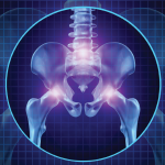
The incidence of articular erosions tends to increase with duration on dialysis. Thus, it is not surprising that our patient, who was on dialysis for 14 years, developed these lesions. Nevertheless, a study by Falbo et al. found only a 40% positive predictive value of radiographic erosions with regard to predicting clinically significant osteodystrophy, highlighting the lack of reliability of using imaging to assess joint pain with this condition.6
Our patient had secondary and tertiary hyperthyroidism. This is characterized by the inability of the kidney to convert vitamin D into its active form, as well as an inability to excrete phosphate, leading to overstimulation of parathyroid hormone. Moreover, the bone-derived hormone fibroblast growth factor-23 (FGF-23) is upregulated due to excessive phosphate retention, also leading to decreased production of active vitamin D. Given these derangements in the elements necessary for proper bone formation, nearly all patients with CKD stage 5 or higher develop some form of osteodystrophy.7
In Sum
Our case serves as an important reminder to consider erosive azotemic osteodystrophy on the differential for hip pain in a patient with long-standing kidney disease, despite radiographic findings that may point in a different direction. On the corollary, erosions are commonly found on imaging studies of CKD patients and do not always correlate clinically with symptoms, as discussed above. Therefore, it is important not to attribute joint pain to renal osteodystrophy simply based on radiographic erosions. Clinicians should further evaluate for an alternative diagnosis.
 Ilana P. Goldberg, MD, is an internal medicine resident at Thomas Jefferson University Hospital, Philadelphia. She obtained her medical degree from Tufts University School of Medicine, Boston.
Samuel Faught, MD, is a rheumatology fellow at Thomas Jefferson University Hospital, Philadelphia.
References
- Fukagawa M, Kazama JJ, Kurokawa K. Renal osteodystrophy and secondary hyperparathyroidism. Nephrol Dial Transplant. 2002;17 Suppl 10:2–5.
- Karchevsky M, Schweitzer ME, Morrison WB, et al. MRI findings of septic arthritis and associated osteomyelitis in adults. AJR Am J Roentgenol. 2004 Jan;182(1):119–122.
- Salam S, Gallagher O, Gossiel F, et al. Diagnostic accuracy of biomarkers and imaging for bone turnover in renal osteodystrophy. J Am Soc Nephrol. 2018 May;29(5):1557–1565.
- Rubin LA, Fam AG, Rubenstein J, et al. Erosive azotemic osteoarthropathy. Arthritis Rheum. 1984 Oct;27(10):1086–1094.
- Lobby MJ, Martel W. Some commonly unrecognized manifestations of metabolic arthropathies. Clin Imaging. 1992 Jan–Mar;16(1):1–14.
- Falbo SE, Sundaram M, Ballal S, et al. Clinical significance of erosive azotemic osteodystrophy: A prospective masked study. Skeletal Radiol. 1999 Feb;28(2):86–89.
- Fang Y, Ginsberg C, Sugatani T, et al. Early chronic kidney disease-mineral bone disorder stimulates vascular calcification. Kidney Int. 2014;85(1):142–150.
