Imaging & Treatment
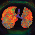
Currently, ILD is primarily detected in patients with CT imaging of the lungs. The pattern found in imaging studies can be significantly helpful in evaluating which underlying connective tissue disease may be at play. Although a non-specific interstitial pneumonia pattern is common in such conditions as systemic sclerosis, systemic lupus erythematosus, myositis and Sjögren’s syndrome, only RA is known to manifest ILD most frequently in a UIP pattern. Such an association is relevant to clinicians because ILD may precede the development of overt clinical signs of an associated connective tissue disease. Thus, ILD in a UIP pattern may herald the onset of RA more so than other related conditions.
On high-resolution CT imaging, UIP classically demonstrates a honeycombing: clustered cystic air spaces of 2–10 mm in diameter with well-defined walls. The affected areas are most commonly located in the subpleural regions and bases of the lungs. In his lecture, Dr. Castellvi recommended that RA patients undergo lung imaging and/or pulmonary function testing at the onset of their disease, regardless of pulmonary symptoms. This approach establishes a baseline for lung involvement and creates a reference point if symptoms develop in the future.
As many rheumatologists are aware, most conventional and biologic disease-modifying anti-rheumatic drugs (DMARDs) used to treat RA can induce alveolar inflammation, interstitial inflammation and/or interstitial fibrosis. Thus, the question of drug-induced lung disease in RA is poignant after a patient starts such a medication.
Methotrexate, one of the most commonly used medications to treat RA, is known to induce hypersensitivity pneumonitis in about 0.43% of patients.3 Typically, this condition occurs within the first year of treatment and is subacute, with a progression of symptoms over days to weeks. Awareness of this phenomenon has resulted in concerns that methotrexate may be associated with an increased incidence or exacerbation of ILD in RA patients.
To further explore this issue, researchers recruited 2,700 patients with early RA and used univariate, multivariate, time-varying and time-to-event Cox proportional hazards analyses to assess methotrexate exposure and other factors that may be associated with a diagnosis of ILD in these patients. The study showed no association in the primary analysis between methotrexate exposure and the incidence of ILD in this population. In fact, methotrexate exposure was associated with significantly less ILD in these patients.4 Additional studies are needed to further evaluate the potential risks or benefits of using various DMARDs in RA patients both with and without active ILD.


