WASHINGTON, D.C.—A mathematical problem that has baffled rheumatologists in recent years is: 10 does not equal 40. This statement refers to the fact that observational studies have estimated that about 10% of patients with rheumatoid arthritis (RA) will go on to develop interstitial lung disease (ILD), but studies that have employed screening high-resolution computed tomography (HRCT) chest imaging have indicated that up to 40% of patients with RA may have subclinical ILD.1 At the ACR Convergence 2024 session Rheumatoid Arthritis-Associated Interstitial Lung Disease: Advances in Screening, Diagnosis and Patient Phenotyping, three outstanding talks shed light on this important subject.
RA-ILD Mortality
The first speaker was Scott Matson, MD, assistant professor, Pulmonary, Critical Care and Sleep Medicine, University of Kansas Medical Center, Kansas City, and he began his talk by noting that the mortality rate is nearly 10% in the first year and 30% by year five in patients with a diagnosis of RA-associated ILD (RA-ILD).2 Even with advancing therapies for RA-ILD (e.g., immunosuppressants and antifibrotic agents), a narrow window exists in which to intervene to significantly reduce mortality. Dr. Matson pointed out that pulmonologists may be hesitant to treat RA-ILD when they have trouble distinguishing it from idiopathic pulmonary fibrosis (IPF). This is because, in 2012, The New England Journal of Medicine published an article demonstrating that patients with IPF treated with a combination of prednisone, azathioprine and N-acetylcysteine were found to have an increased risk of death and hospitalization compared with patients treated with placebo.3 This has forced clinicians to be very careful in distinguishing RA-ILD from IPF to determine if immunosuppression may be helpful or harmful.
Dr. Matson went on to describe in detail a study from Solomon et al., a prospective, randomized controlled trial that exclusively enrolled RA-ILD patients. In this phase 2 study, more than 120 patients with RA-ILD were assigned to receive pirfenidone, an antifibrotic agent, vs. 60 patients assigned to the placebo arm. The trial was stopped early due to slow recruitment and the COVID-19 pandemic, but pirfenidone did appear to slow the decline in the rate of forced vital capacity (FVC) over time in patients with RA-ILD.4 This is a similar result to that seen in other studies of antifibrotic medications, with an observable slowing of the FVC reduction over time. Dr. Matson explained that, given these results, patients with RA-ILD who demonstrate a predominantly usual interstitial pneumonia (UIP) pattern would typically be recommended for antifibrotic treatment. As to the best method for immunosuppression in these patients, the jury is still out and more data are needed.
Mining Electronic Medical Records
The second speaker, Bryant England, MD, PhD, associate professor in the Division of Rheumatology, University of Nebraska Medical Center, Omaha, began his talk by discussing the issue of wide variability in estimates of RA-ILD incidence and prevalence. Such variations raise questions: Are we over- or underdiagnosing RA-ILD? How wide is the spectrum of RA-ILD disease? One topic on which general agreement exists is that clinicians must seek to identify RA-ILD as early as possible to have any hope of reducing mortality (of note, the median survival for patients with RA-ILD is 3–10 years).5
Dr. England pointed out that electronic medical record systems contain a wealth of clinical information, and, using the correct algorithmic techniques, this data could be harnessed to better identify patients with RA-ILD. In a 2020 study on this matter, Dr. England et al. used the records of more than 500 patients with RA from the Veterans Affairs administrative data sets and identified patients with RA-ILD based on International Classification of Diseases Ninth Revision (ICD-9) and Tenth Revision (ICD-10) codes. They then characterized the performance of administrative algorithms for identifying RA-ILD compared with doing so by a detailed chart review. The authors found that the best performing algorithm was the one that incorporated at least two ILD diagnosis codes 30 days apart, a pulmonologist diagnosis or CT/pulmonary function test (PFT) evidence of ILD prior to the rheumatologist’s diagnosis and exclusion of other ILD causes.6
In a separate study, Dr. England et al. furthered their work on RA-ILD algorithms by applying text mining techniques to HRCT reports. In this study, patients with RA-ILD were identified and ILD-related terms, such as reticulation, ground glass, honeycomb and interstitial, were identified in CT chest reports by natural language processing software. Importantly, the researchers made sure to confirm that no negating terms were around these words (i.e., such phrases as no honeycombing or absence of reticulations). By combining ILD-related terms from CT reports with administrative algorithms, the positive predictive value for identifying RA-ILD exceeded 90%.7 Projects like these are important to think about identifying RA-ILD in real-world scenarios; frequently, patients with RA present to their primary care doctor with cough or other vague pulmonary symptoms and undergo CT chest imaging. Dr. England asked the audience to picture this scenario and add the element of an artificial intelligence system working in the background, culling radiology reports from such patients and flagging any with potential concern for RA-ILD, prompting the treating rheumatologist to order PFTs and consider a pulmonology referral.
Nailfold Capillaroscopy
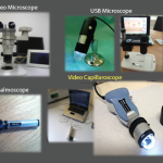
The final speaker was Vanessa Smith, MD, PhD, associate professor of rheumatology and head of clinics, Ghent University Hospital, Belgium, who focused on the topic of nailfold capillaroscopy. At first glance, nailfold capillaroscopy and RA-ILD may not seem to fit together. However, the purpose of Dr. Smith’s talk was to remind the audience of how important the nailfold exam is in all patients with concern for ILD to identify scleroderma-spectrum diseases. In 1973, Maricq and LeRoy published a landmark study on the use of wide field microscopy to identify patterns of abnormal nailfold capillaries in patients with connective tissue diseases.8 Cutolo further expanded on this work in later years by employing 200x magnification techniques to look at nailfold capillaries in even greater detail. In 2020, Dr. Smith joined Dr. Cutolo and others in writing a consensus paper on nailfold capillaroscopy in patients with Raynaud’s and systemic sclerosis. In this paper, the authors were able to describe nailfold density, dimension, morphology and hemorrhage to create two categories: scleroderma pattern and non-scleroderma pattern.9
The fact that only patients with scleroderma-spectrum diseases should have a scleroderma pattern on nailfold capillaroscopy is helpful when evaluating for the underlying cause of a patient’s ILD. Dr. Smith provided a clinical example in which a patient presented with ILD in a nonspecific interstitial pneumonia (NSIP) pattern with concomitant inflammatory arthritis. The initial impulse of the treating provider was to label the patient as possibly having RA-ILD, but the nailfold capillaroscopy demonstrated a scleroderma pattern. Upon further evaluation, this patient was found to have antisynthetase syndrome as the cause of his disease. In a different case, a young woman was identified as having ILD in a UIP pattern and her lab studies showed a range of low titer antibodies in a confusing pattern. Her nailfold capillaroscopy was normal, and ultimately she was diagnosed as having a genetic cause for her ILD. The main message conveyed in these cases is that the absence or presence of scleroderma pattern nailfold capillaroscopy results can aid a great deal in finding the true etiology of ILD in each patient.
In Sum
The talk highlighted an unmet need: correctly identifying and treating patients with RA-ILD early in their disease course. Achieving success in this area is challenging, and a collaboration between rheumatologists and pulmonologists is important to improve the status quo and better patient care.
Jason Liebowitz, MD, is an assistant professor of medicine in the Division of Rheumatology at Columbia University Vagelos College of Physicians and Surgeons, New York.
References
- Juge PA, Granger B, Debray MP, et al. A risk score to detect subclinical rheumatoid arthritis–associated interstitial lung disease. Arthritis Rheumatol. 2022 Nov;74(11):1755–1765.
- Farquhar HJ, Beckert N, Beckert L, et al. Survival of adults with rheumatoid arthritis associated interstitial lung disease—A systematic review and meta-analysis. Semin Arthritis Rheum. 2023 Jun;60:152187.
- Idiopathic Pulmonary Fibrosis Clinical Research Network; Raghu G, Anstrom KJ, et al. Prednisone, azathioprine, and N-acetylcysteine for pulmonary fibrosis. N Engl J Med. 2012 May 24;366(21):1968–1977.
- Solomon JJ, Danoff SK, Woodhead FA, Et al. Safety, tolerability, and efficacy of pirfenidone in patients with rheumatoid arthritis-associated interstitial lung disease: A randomised, double-blind, placebo-controlled, phase 2 study. Lancet Respir Med. 2023 Jan;11(1):87–96.
- Juge PA, Wemeau L, Ottaviani S, et al. Increased mortality in patients with RA-associated interstitial lung disease: Data from a French administrative healthcare database. RMD Open. 2023 Dec 1;9(4):e003491. Erratum in: RMD Open. 2024 Jan 30;10(1):e003491corr1.
- England BR, Roul P, Mahajan TD, et al. Performance of administrative algorithms to identify interstitial lung disease in rheumatoid arthritis. Arthritis Care Res (Hoboken). 2020 Oct;72(10):1392–1403.
- Luedders BA, Cope BJ, Hershberger D, et al. Enhancing the identification of rheumatoid arthritis-associated interstitial lung disease through text mining of chest computerized tomography reports. Semin Arthritis Rheum. 2023 Jun;60:152204.
- Maricq HR, LeRoy EC. Patterns of finger capillary abnormalities in connective tissue disease by “wide-field” microscopy. Arthritis Rheum. 1973 Sep–Oct;16(5):619–628.
- Smith V, Herrick AL, Ingegnoli F, et al. Standardisation of nailfold capillaroscopy for the assessment of patients with Raynaud’s phenomenon and systemic sclerosis. Autoimmun Rev. 2020 Mar;19(3):102458.