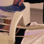
Shidlovski / SHUTTERSTOCK.COM
With the aid of increasingly sophisticated neuroimaging technology, research into how the brain activates and changes in patients with chronic pain is delivering fascinating information that will hopefully pave the way to tailored, individual treatment of chronic pain.
Over the past several years, data from neuroimaging studies have provided a new understanding of what occurs in the brains of people with chronic pain conditions such as fibromyalgia and osteoarthritis. Recent data, for example, show that increased processing of pain in specific areas of the brain associated with the expectation of pain (e.g., the insular cortex) correlated with the extent and severity of pain in patients with fibromyalgia and osteoarthritis.1
These studies are helping researchers better understand the complex interplay of changes in brain pathways that affect both the sensory and emotional aspects of pain, and how changes in these two pathways interact to create the perception and experience of chronic pain. This is described well in a review of the current research on pain neuroimaging by researchers from the United Kingdom, who detail the way in which chronic pain appears to alter brain function and structure in these patients.2 Specifically, functional imaging studies show that two primary pain pathways, the lateral and medial pain pathways, are involved in what has become known as the pain matrix. Researchers think these two pathways account for the sensory aspects of pain (i.e., the lateral pathway) as well as the emotional aspects of pain (i.e., the medial pathway).2

Professor Jones
Anthony Jones, MD, professor of neuro-rheumatology at the University of Manchester in England and senior author of the review, describes it as the need to integrate the two important pathways in the brain that tells a person they’re experiencing pain: the hard-wired information from damaged pain fibers in the body (i.e., the sensory pathway), and the activation of brain areas associated with emotion, attention and anticipation (i.e., the emotional pathway) that conditions the brain to expect the experience of pain based on prior pain experiences. That latter point is critical to understanding that even the subjective experience of chronic pain, so baffling to clinicians and patients, is being shown by neuroimaging studies to also be based in physiological changes in the brain and how the brain adapts to or accommodates acute pain experiences.
“In different patients, different things will drive the pain that’re often not related to physical damage,” Professor Jones says.
Despite a growing recognition and appreciation for the neurobiology of chronic pain, ongoing studies struggle to find specific and unique pain biomarkers, according to Professor Jones. “This is still very much an evolving field of research,” he says.
Recent Research

Dr. Harris
Among the more recent research in this field is a study published by researchers at the University of Michigan that examined whether brain structures or connectivity patterns associated with widespread pain could be identified on neuroimaging.3 “We and others have noticed that many patients with localized pain conditions, such as knee osteoarthritis and pelvic pain, have pain in locations other than where their primary complaint is,” says the study’s senior author, Richard Harris, PhD, associated professor of Anesthesiology and Rheumatology at Michigan Medicine in Ann Arbor, Mich.
Using data from 1,079 participants in the Multidisciplinary Approach to the Study of Chronic Pelvic Pain Research Network (MAPP) study, Harris and colleagues compared three cohorts: patients with a clinical diagnosis of urological chronic pelvic pain syndrome (UCPPS), a pain-free control group and patients with fibromyalgia. All participants completed questionnaires on pain severity, function and pain location. A subset also underwent functional and structural magnetic resonance imaging.
The study found that some of the patients with UCPPS had pain beyond the pelvic area, and that those with widespread pain showed changes in brain structure and function identical to the changes seen in the fibromyalgia. Specifically, widespread pain in both the UCPPS and fibromyalgia patients was associated with increases in brain gray matter and connectivity in sensory encoding brain areas.
The implications of these findings point to what researchers hope they can reap from this entire line of investigation. “It’s important to realize that a person who presents with localized pain may also have a more broad body pain presentation that may respond better to centrally acting interventions, as opposed to treatment focused on the local complaint,” says Dr. Harris.
Yes, It’s All in Their Head
This research and ongoing investigation shows chronic pain is indeed all in the patient’s head—specifically, their brain. “The question often is, ‘So the pain is all in my head, is it?’ to which the answer is yes, it must be, because that’s where the brain is, and pain can only be experienced as a result of activity in the brain,” says Professor Jones. This once meant a dismissal of a patient’s subjective experience, but the evolving understanding of pain as a brain problem now means neuroimaging can capture pain pathways, thereby possibly allowing doctors to devise treatment measures that better ease both the physical and emotional discomfort of pain than traditional measures.
For Professor Jones, this means an approach to pain management that harnesses the brain to correct itself. “The brain is a powerful organ and can be trained to take more control over the experience of pain,” he says. In a recently published study, he and his colleagues showed the ability of mindfulness-based talking therapies to substantially reverse some of the “fine-tuning” problems of a brain trying to integrate both the sensory and emotional pathways turned on by pain.4 The study showed that patients, through mindfulness-based talking therapies, can learn to take more control over their pain experience, according to Professor Jones.
Along with talking therapies, he emphasized the important role of exercise in managing chronic pain. “Long-term exercise and talking therapies have the best ratio of benefit to harm,” he says. “So these are worth investing in in the long term.”
Going Forward

Dr. Davis
As studies continue to try to identify specific biomarkers of chronic pain, discussion of the best ways to apply this research is fully underway. In September 2017, a presidential task force of the International Association for the Study of Pain published a consensus statement recommending against the use of brain imaging as a test for chronic pain.5 Led by Karen Davis, PhD, head of the division of Brain, Imaging and Behaviour-Systems Neuroscience at the Krembil Research Institute in Toronto Western Hospital in Toronto, Ontario, Canada, the consensus statement discusses the medical, legal, and ethical issues involved with using brain imaging tests for chronic pain.
Dr. Davis says that although the task force believes brain imaging can serve an important role in discovering the brain mechanisms underlying pain, and possibility help predict therapies that help individual patients, she cautioned that some negative unintended applications of such imaging could disadvantage the patient. “A key concern of using biomarkers of pain regards diagnostics of chronic pain, whether an individual patient does or doesn’t experience pain,” she says, adding that such diagnostics “then could be used to deny or restrict medical care or coverage of costs.”
Professor Jones agrees that until a clearer understanding of the neurobiology of pain emerges and biomarkers are identified, the question of how to use them in clinical practice remains a question. Functional brain imaging “has no place in the routine management at the moment,” he says.
Mary Beth Nierengarten is a freelance medical journalist based in Minneapolis.
References
- Brown CA, El-Deredy W, Jones AK. When the brain expects pain: Common neural responses to pain anticipation are related to clinical pain and distress in fibromyalgia and osteoarthritis. Eur J Neurosci. 2014 Feb;39(4):663–672.
- Morton DL, Sandhu JS, Jones AK. Brain imaging of pain: State of the art. J Pain Res. 2016 Sep 8;9:613–624.
- Kutch JJ, Ichesco E, Hampson JP, et al. Brain signature and functional impact of centralized pain: A multidisciplinary approach to the study of chronic pelvic pain (MAAP) network study. Pain. 2017 Oct;158(10):1979–1991.
- Jones AKP, Brown CA. Predictive mechanisms linking brain opioids to chronic pain vulnerability and resilience. Br J Pharmacol. 2017 Apr 27.
- Davis KD, Flor H, Greely HT, et al. Brain imaging tests for chronic pain: Medical, legal and ethical issues and recommendations. Nat Rev Neurol. 2017 Oct;13(10):624–638.
