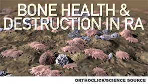
Osteoclasts are the bone-resorptive cells that play the key role in physiological skeleton development and homeostatic bone remodeling. Consequently, abnormal osteoclast function can lead to osteoporosis, rheumatoid arthritis (RA) and other diseases. Investigators are, thus, eager to learn what makes an osteoclast healthy and functioning and what can go wrong, thereby creating pathology. A new study provides a novel contribution to our understanding of osteoclast physiology: RBP-J is a key regulator of osteoclastogenesis and bone remodeling.
RBP-J is also known as CSL and CBF1. It is the recombinant recognition sequence binding protein at the Jk site. It is known to function as a central transcription factor, receiving signals from multiple signaling pathways. We now know that it also regulates bone remodeling. What makes the finding especially interesting is that the newly discovered RBP-J-centered signaling pathway appears to contribute to the development of the inflammatory bone erosion that is a hallmark of RA. The pathway also explains the mystifying results of a previous genome-wide association study that linked a variant in RBP-J with the development of RA.
The current research was performed by Baohong Zhao, PhD, assistant scientist in the Arthritis and Tissue Degeneration Program at the Hospital for Special Surgery in New York City and colleagues and published online Oct. 20 in the Journal of Clinical Investigation.1 In their paper, the investigators not only describe the signaling pathway, but also describe how environmental cues that modulate expression/function of the transcription factor RBP-J appear to influence osteoclast differentiation and bone remodeling.
A new study provides a novel contribution to our understanding of osteoclast physiology: RBP-J is a key regulator of osteoclastogenesis & bone remodeling.
What Has Previously Been Shown
Osteoclastogenesis begins with RANK signaling in osteoclast precursors, as well as costimulation from immunoreceptor tyrosine-based activation motif-containing (ITAM-containing) receptors/adaptors. The two main ITAM-containing tyrosine kinase binding proteins involved in this process are DNAX-activating protein 12 (DAP12) and Fc receptor common ɣ subunit (FcRɣ). These ITAM-adaptors are known to associate with various receptors and mediate signaling. Recent work has also revealed that the RANK- and ITAM-signaling pathways are integrated by a multiprotein signaling complex comprising BTK/TEC/BLNK/SLP76/PLCɣ2. Up until now, however, investigators have not known how the costimulatory signals are regulated and integrated with RANK signaling.
Where RBP-J Fits In
RBP-J controls excessive bone erosion through facilitation of RANK signaling. The ligand for RANK (RANKL) induces osteoclastogenesis, but to do this, it first requires calcium signaling to activate NFATc1. NFATc1 is established as the master regulator of osteoclastogenesis, and its activation requires sustained calcium signaling, as opposed to an acute and transient calcium flux. RBP-J provides sustained calcium signaling through the suppression of calcium fluctuations via a TGF-β/PLCɣ2/calcium axis. Moreover, after RANKL stimulation during osteoclastogenesis, RBP-J expression decreases.
Thus, RBP-J imposes the requirement for ITAM-mediated costimulation of RANKL. It also inhibits ITAM-mediated expression and function of the previously identified member of the key signaling complex, PLCɣ2, as well as activation of downstream calcium-CaMKK/PYK2 signaling. It does this by suppressing induction of key osteoclastogenic factors, such as NFATc1, BLIMP1 and c-FOS. RBP-J thereby limits the crosstalk between ITAM and RANK/TNFR and allows for fine-tuning of osteoblastogenesis during bone homeostasis. This is the process that appears to underlie bone health, as well as bone destruction seen in RA.
Experimental Details
In order to elucidate this pathway, the researchers used powerful, high-tech, next generation, whole transcriptome sequencing. They began their research with two mouse models of osteoporosis (Dap12-/- and Dap12-/-Fcrg-/-). Their experiments revealed that the osteoporotic bone phenotype in these mice is mediated by RBJ-1. Specifically, deletion of Rbpj in these mice substantially rescued the defects in bone remodeling, thereby reversing the osteoporotic bone phenotype. A more detailed analysis revealed that knocking out Rbpj bypassed the requirement for ITAM-mediated costimulation of RANKL-induced osteoblastogenesis.
The investigators then looked more closely at the role of PLCɣ2 in transgenic and control mice. Its relative expression and phosphorylation in the different strains suggested that it plays an important regulatory role in the pathway. In contrast, the previously identified ITAM-binding protein FcRɣ did not appear to significantly affect bone remodeling. Inhibition of PLCɣ2 activity significantly reduced the enhanced RANKL-induced osteoclastogenesis in RBP-J-deficient cells. A more detailed analysis also revealed that RBP-J regulates TGF-β signaling and, thus, suppresses the expression and activity of basal PLCɣ2 and downstream calcium-CaMKK-PYK2 signaling. Investigators additionally found that the RBP-J target gene Tgfbr1 (which codes for TGF-β receptor I) was expressed at a rate that was 2.5 times higher in RBP-J-deficient osteoclast precursors compared to controls.
The team then turned to a different mouse model of bone disease: a TNF-α-induced model of inflammatory bone resorption. They found that RBP-J deficiency allows TNF-α to induce osteoclast formation and bone resorption in DAP12-deficient mice. Moreover, RBP-J deficiency in the absence of ITAM-mediated costimulatory signaling enabled TNF-α to induce NFATc1 and BLIMP1 expression and osteoclastogenesis. Thus, RBP-J also imposes the requirement for ITAM-mediated costimulation in TNF-α-induced osteoclastogenesis.
Lara C. Pullen, PhD, is a medical writer based in the Chicago area.
Reference
- Li S, Miller CH, Giannopoulou E, Hu X, Ivashkiv LB, Zhao B. RBP-J imposes a requirement for ITAM-mediated costimulation of osteoclastogenesis. J Clin Invest. 2014 Nov 3;124(11):5057–5073.