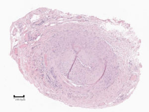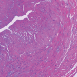
Figure 1: Active arteritis. Inflammatory cells are present in the media in the lower left aspect of the vessel wall. (Click to enlarge.)
At the end of 2023, the Society for Cardiovascular Pathology (SCVP) published consensus guidelines on the diagnostic approach to temporal artery biopsy.1 Through this publication, SCVP hopes to bring more uniformity to the processing, interpretation and reporting of these specimens, taking into consideration the most up-to-date literature available. These guidelines have obvious impact on clinical practice, particularly in the diagnosis and, thus, management of giant cell arteritis (GCA). We wish to highlight some of the important messages from our consensus document and engage the rheumatology community in synthesizing such guidelines into future, unified diagnostic criteria.
Gold Standard for GCA Diagnosis
Although imaging criteria continue to improve, the current standard against which the sensitivity and specificity of those studies is measured is biopsyproven disease. The 2021 ACR/Vasculitis Foundation Guideline speaks to recommendations specifically for the use of biopsy in diagnosing GCA, although it acknowledges the recommendations are conditional and with low level of evidence.2
We acknowledge the broader, and reasonable, recommendations in the 2022 ACR/EULAR classification criteria.3 Nonetheless, we (admittedly somewhat self-servingly) support the recommendation of continuing to use biopsy as a gold standard at a minimum until higher quality evidence for other diagnostic techniques or algorithms is available.
The imperfect sensitivity of temporal arterial biopsy for GCA (currently estimated at 61–77%), given the spatial and temporal heterogeneity of the disease, however, is acknowledged.4,5 In cases of high clinical suspicion and negative biopsy, we obviously support the utility of imaging and other diagnostic modalities.
Healed Injury Is Not Specific for Arteritis

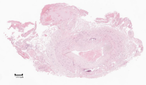
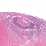
Figure 2: Healed arterial injury. There is intimal hyperplasia, adventitial fibrosis, and calcification of the internal elastic lamina (Monckeberg’s medial calcification), but no inflammation. (Click to enlarge.)
“Healed arteritis” has received much attention in the literature over the years, with a number of criteria proposed, but without certainty as to the clinical significance. The latest evidence available to us at the time we wrote our guidelines, however, demonstrates that all the findings of healed arteritis can be seen in cases without GCA or any other vasculitis.
Histomorphologically, there is overlap between trauma (accidental, iatrogenic, prior biopsy), age-related changes and healed arteritis. Hence we specifically recommend the diagnostic term “healed arterial injury” to reflect that something damaged the temporal artery, but we cannot comment as to the etiology. This is an important recognition in developing future diagnostic algorithms because the degree to which post-test probability is informed by “healed arteritis” is insufficiently studied.
Not Every Inflamed Temporal Artery Is Due to GCA
An important section of our consensus document speaks to the differential diagnosis of active arteritis in a temporal artery biopsy. We acknowledge that most such cases will represent GCA, especially in the context of high clinical suspicion (pre-test probability) and the marked prevalence of GCA compared with the other conditions, especially in the temporal artery. That said, especially for unusual presentations of disease, the consideration of other diagnoses remains valuable.
Synoptic Reporting & Scoring?
Our consensus presents recommended report formats but stops short of an overly synoptic report. The use of such reporting templates is prevalent in cancer diagnostics, but is not yet mature in non-neoplastic diagnoses. We know that every patient has a unique case of the disease, and as much as we lump these into GCA, personalized medicine dictates we go deeper. A uniform terminology sets the groundwork for developing such synoptic reports, and those may inform multi-modality diagnostic criteria, scoring of disease, choice of therapeutic approach, enrollment in clinical trials and evolution of future guidelines from our respective societies—or even combined guidelines.
We Need to Work Together
Please involve pathologists in your multidisciplinary discussions for GCA and any other tissue-based diagnosis. Communication and partnership are key to evolving understanding in these diseases.
Moayad Alqazlan, MBBS, is a cardiovascular pathology fellow in the Department of Laboratory Medicine and Pathobiology, University of Toronto, Ontario.
Vidhya Nair, MBBS, MD, is chair of the Department of Pathology and Laboratory Medicine, University of Ottawa, Ontario.
Michael A. Seidman, MD, PhD, is director of quality and safety in surgical pathology, director of autopsy, and staff pathologist at the University Health Network Laboratory Medicine Program at Toronto General Hospital. He is an assistant professor in the Department of Laboratory Medicine and Pathobiology, University of Toronto, Ontario.
References
- Nair V, Fishbein GA, Padera R, et al. Consensus statement on the processing, interpretation and reporting of temporal artery biopsy for arteritis. Cardiovasc Pathol. 2023 Nov–Dec;67:107574.
- Maz M, Chung SA, Abril A, et al. 2021 American College of Rheumatology/Vasculitis Foundation Guideline for the management of giant cell arteritis and Takayasu arteritis. Arthritis Rheumatol. 2021 Aug;73(8):1349–1365.
- Ponte C, Grayson PC, Robson JC, et al. 2022 American College of Rheumatology/EULAR classification criteria for giant cell arteritis. Arthritis Rheumatol. 2022 Dec;74(12):1881–1889.
- Rubenstein E, Maldini C, Gonzalez-Chiappe S, Chevret S, Mahr A. Sensitivity of temporal artery biopsy in the diagnosis of giant cell arteritis: A systematic literature review and meta-analysis. Rheumatology (Oxford). 2020 May 1;59(5):1011–1020.
- Dua AB, Husainat NM, Kalot MA, et al. Giant cell arteritis: A systematic review and meta-analysis of test accuracy and benefits and harms of common treatments. ACR Open Rheumatol. 2021 Jul;3(7):429–441.


