The Case
A 64-year-old man with history of type-II diabetes (well controlled on sitagliptin/metformin), hypertension, and dyslipidemia presents with complaints of an increasingly painful left lower-extremity lesion present for two to three months, which has increased in size and become “firm and stiff,” per the patient’s report. He does remember local trauma to the area prior to the onset of the lesion. He is afebrile and feels otherwise well, and an extensive review of systems is negative.
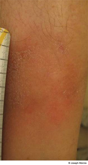
A full-body skin examination reveals the lesion seen in Figure 1, located on the medial aspect of the left lower extremity only, above the medial malleolus. Scattered venous varicosities are present throughout the lower extremity as well as trace-1+ pitting edema to the bilateral ankles. The remainder of the patient’s physical exam was unremarkable.
What is your diagnosis?
- Lipodermatosclerosis
- Morphea
- Cellulitis
- Necrobiosis lipoidica diabeticorum
- Limited systemic sclerosis

