“Anything except prednisone,” her voice sharpened. “I once had an asthma attack, and the emergency room doctor gave me prednisone. I couldn’t sleep for a week, and my blood sugars skyrocketed. And in case you didn’t notice, I have osteoporosis. My bones are brittle enough without adding prednisone, thank you.”
Test Results Start Coming In
I ate lunch on the fly that day and immersed myself in the afternoon’s schedule of stable—and not-so-stable—patients. (I tapped a middle-aged man’s hot, swollen ankle and found gout crystals. Back-to-back lupus patients were doing well, a first for both of them. A rheumatoid patient was flaring, and we discussed adding a biologic, infliximab, to her background weekly methotrexate. An elderly man with unexplained headaches and muscle stiffness came in for his first visit after a biopsy of the temporal artery confirmed the diagnosis of giant cell arteritis.)
Late in the afternoon, I noticed the lab tech had paper-clipped Mrs. N’s lab results onto her chart and placed it squarely in the center of my desk. I sat down and finished off my apple and opened a bag of Fritos. Circled in red were two particularly worrisome results: a C-reactive protein level of 19.5 mg/dL (normal: <0.8 mg/dL) and an erythrocyte sedimentation rate (ESR) of 110 (normal: <20). The CRP and ESR reflected extraordinarily high levels of inflammation, but didn’t clarify if this was due to a persistent infection, an immunologic disorder or an underlying malignancy. The results did, however, lay to rest any notion that Mrs. N was on the tail-end of a self-limited illness.
I pulled Joanne out of an adjacent exam room. “Can you call the medical center and set up an echocardiogram for Mrs. N?” Endocarditis, a life-threatening bacterial infection of a heart valve, would fit with Mrs. N’s clinical presentation. I ran my finger down the order sheet. Okay, good, I ordered blood cultures. I opened the next chart, but my mind refused to move on. Admit Mrs. N? Continue an outpatient work-up? I processed the available information—her healthy appearance, stable vitals and normal white blood count and kidney function—and decided to keep Mrs. N out of the hospital. For now.
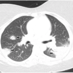
Thankfully, Mrs. N’s blood cultures were sterile. The echocardiogram demonstrated only a slightly stiff aortic valve, but no evidence of seeding by bacteria. At her follow-up visit, she had lost three more pounds but continued to look well. I reviewed the unremarkable CT scan of the chest, an important negative, because lymph node enlargement or tumors can often be missed on a routine chest X-ray. On her lab, a normal CPK and aldolase and an anti-nuclear antibody (ANA) in the normal range made a diagnosis of myositis or lupus unlikely. Likewise, with an absent anti-neutrophil cytoplasmic antibody (ANCA) and a normal urinalysis, several rare forms of vasculitis had been ruled out.


