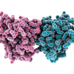
One way to address this concern is to identify potential endogenous TLR ligands by their ability to directly bind TLR2 or TLR4. We employed the extracellular domain of TLR2 and TLR4 to pull down potential ligands from macrophage cell lysates. With this technique, we identified an endoplasmic reticulum expressed chaperone protein, gp96.23 Other studies identified gp96 as a master chaperone responsible for targeting TLRs to either the endosome or the cell surface.24 In the absence of gp96 in macrophages, the TLRs are expressed but do not localize to their respective locations and are not functional. We observed that gp96 binds to TLR2 and that it is highly expressed in RA synovial tissue and, importantly, extracellularly in RA synovial fluid. The gp96 in the RA synovial fluid is significantly increased compared to fluids from patients with psoriatic arthritis, ankylosing spondylitis, or osteoarthritis.
Using a highly purified, ultralow endotoxin gp96, we were able to demonstrate that gp96 was a potent activator of macrophages, inducing inflammatory cytokines. Additionally, the induction of inflammatory cytokines from RA synovial fluid macrophages by gp96 was significantly greater than observed with peripheral blood monocytes. Further, the concentration of gp96 in the RA synovial fluid was strongly correlated with the expression of TLR2 on RA synovial fluid macrophages, suggesting a self-perpetuating process. Supporting a role for endogenous TLR2 ligands in the progression and joint destruction that may be observed in RA, TLR2 is highly expressed in the leading edge of the RA pannus as it erodes into bone, suggesting that constituents of the bone may contribute to the inflammatory process.25 Consistent with the role of TLR2 in the pathogenesis of RA, incubation of RA synovial tissue explants with a neutralizing antibody to TLR2, suppresses the constitutive expression of TNF-α, suggesting a role for endogenous TLR2 ligands in the ongoing inflammation.26 Further supporting a role for gp96 in RA, our data demonstrate that RA synovial fluid gp96 is capable of activating macrophages mediated through TLR2, and that RA synovial fluid macrophages express significantly increased levels of cell surface gp96, which is capable of activating adjacent macrophages, presumably through TLR2.27 These observations suggest a self-perpetuating cycle, where inflammation results in the increased expression and release of gp96, which then results in further TLR-mediated macrophage activation.
In an attempt to identify other endogenous TLR2 ligands in the synovial tissue of patients with RA, we went fishing, employing a yeast two-hybrid system in which the extracellular domain of TLR2 was the bait, and proteins expressed by a cDNA library constructed from the synovial tissue of patients with longstanding and erosive, active RA were used as the prey. The molecule that most frequently interacted with TLR2 was Snapin, a small molecular-weight protein that is critical for neurotransmission and the transport of autophagosomes within neurons.28 After confirming that Snapin bound to TLR2, we demonstrated that Snapin was capable of activating through TLR2. Snapin is highly expressed in RA synovial tissue examined by immunohistochemistry and qRT-PCR.29 There was a significant correlation between Snapin in the RA synovial tissue by qRT-PCR and clinical disease activity determined by number of swollen joints. Supporting the relevance of Snapin to the pathogenesis of RA, a neutralizing antibody to Snapin suppressed the ability of some RA synovial fluids to activate macrophages. These observations identify gp96 and Snapin as endogenous TLR2 ligands that contribute to the pathogenesis of RA.

