Pediatric rheumatologists, I would argue, are often cut from a similar cloth. We are generally creatures of the clinic, finding safety and security in our ability to navigate through a day of patients that we have often known for years. In many cases, we’ve watched our patients grow up, and we’ve established deep bonds with them and their families, guiding them from a time of often-critical illness toward health. We see our share of consults on the floors, but they come at the end of a clinic day and we can usually count them on one hand per day. And so there was a sense of unease when I found myself making a patient list to keep track of our new admissions and continuing inpatients that numbered more than 20.
Beginning in late April, we (my partner, Alexis Boneparth, MD, and I) began to see a new phenomenon: difficult to understand at first and difficult to separate from the more typical (if such a word can be used today) and scattered cases of cytokine storm associated with COVID-19 infection.
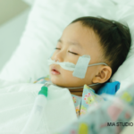
These patients were different. Often coming in hypotensive, with fever and rash, they quickly found themselves in the intensive care unit (ICU). We heard of them from our gastroenterologist colleagues, who had received them from surgeons after computerized tomography (CT) scans of the abdomen suggested not appendicitis, but a raging inflammatory colitis. We read notes from otolaryngologists describing a pharyngitis so severe it resulted in frank trismus, and from neurologists regarding severe headaches and nuchal rigidity that warranted a lumbar puncture.
Slowly, a picture began to form: A new syndrome, completely different from the original infectious process, was being born in our emergency department.
Observations
This syndrome could be differentiated from COVID-19 and subsequent cytokine storm by the absence of acute respiratory distress syndrome (ARDS) and hemophagocytic lymphohistiocytosis (HLH). Not one of our patients (approximately 30 at that point in time) had displayed either of these findings. Further, the syndrome appeared to be defined by three major features found in most of the cases: prolonged and high fever; severe abdominal pain; and, bizarrely, a robust clinical myocarditis that resulted in serum concentrations of brain natriuretic peptide (ntBNP) in the many tens of thousands. Associated with this presumed myocarditis was a functional defect in ejection fraction in about two-thirds of the patients.
As we learned more, we saw that about half of the patients presented in shock—in some cases severe shock, with blood pressures only barely able to be measured—out of proportion to the moderate cardiac dysfunction seen. Alongside these features, we saw nonspecific rashes and, sometimes, mucosal inflammation of the oral cavity or, perhaps, a conjunctivitis.
The requests began to come for our thoughts as consultants. The question was raised: Is this Kawasaki disease? Kawasaki disease is both a disease of childhood, known well to pediatricians, and an autoimmune process. It presents with fever, rash, and other findings. It ends, in some cases despite the best treatments, with coronary aneurysms caused by a necrotizing vasculitis of coronary vessels.
This new phenomenon has some similarities—sustained, very high fever; ill-appearing children; and skin and mucosal changes. Some patients present with abdominal pain and shock, which can also be seen in severe forms of Kawasaki. And yet, at the same time, we had not seen true vasculitis, and the degree of illness, along with the additional unusual features, suggest that calling this Kawasaki disease would be a mischaracterization. So we called it something else—and variants of this name were described by others as well—a multi-system inflammatory syndrome, COVID-associated.
CLINICAL GUIDANCE AVAILABLE
The ACR has issued a new clinical guidance document for the management of Multisystem Inflammatory Syndrome in Children (MIS-C) Associated with SARS-CoV-2 & Hyperinflammation in COVID-19.
Care
Sensing we would be shortly assaulted by many more of these patients, our institution (Columbia University Irving Medical Center/Morgan Stanley Children’s Hospital) began to create a plan to coordinate how we would manage this disease, especially in its initial, potentially disastrous phases. Along with our colleagues in pediatric infectious disease and pediatric intensive care, we drew up a set of guidelines to tackle patients in their many different presentations.
We focused on ensuring we identified and properly treated those patients in shock, and proposed to use potent therapy—steroid pulses and, for many patients, intravenous immunoglobulin (IVIG). We also considered the possibility of adding interleukin 1 (IL-1) blockade based on its theoretical basis in Kawasaki disease. For those not in shock, we could tailor a more gentle therapy of low-dose steroids. Our colleagues, usually hesitant in allowing us to use the traditional rheumatologist’s intervention of pulse steroids, gave us surprising leeway.
Within a week, the patients began to arrive. At first, three or four a day, and then over one weekend we were inundated by 20 patients in a three-day span. During that week and weekend, I would wake up, make sure I played teacher for at least an hour to my children (who were refusing to do any kind of schoolwork) and then drive down the empty highways into New York, usually answering two or three phone calls on the way, and remain at work until after midnight.
Our protocol was printed up and distributed across the hospital. We saw patients in the emergency department or as they arrived in the ICU or on the floors, and encouraged treatment with the therapies we use so often in our rheumatology patients.
The first of many moments that I will remember for years came when I overheard a nurse expressing her amazement at “how well those steroids worked.” The patient she had in mind was on such heavy doses of dopamine that one could see “his heart beating through his chest”; within an hour of a steroid pulse, he was able to begin the process of weaning the dopamine, and quickly and effortlessly completed the wean over the next 24 hours.
Often, I would see patients critically ill and then, only 12 hours later, they would be sitting on their beds, nonchalantly playing on their parents’ iPads.
What I had not been sure of was the question of aneurysms and vasculitis. Would we see them? I was skeptical. Could this truly be a vasculitis? That was what, in many ways, defined Kawasaki disease for me—the presence of vascular inflammation and destruction. Perhaps ironically, I had come to Columbia to study just this process, having spent the previous four years working on developing and manipulating a mouse model of this same disease. I had spent many afternoons and evenings looking at sections of mouse hearts with obliterated vessels. I had read many, many journal articles on the theoretical basis for how these aneurysms formed—from the neutrophils digesting the vessels into the adventitia, to the myofibroblasts proliferating and causing neointimal hyperplasia and stenosis. To think this process would be occurring in a pandemic, in front of my eyes, seemed simply too unlikely a bet.
And then it happened. On May 12, one of our earliest patients had a follow-up ultrasound, around 12 days after his illness, and demonstrated a moderate-sized aneurysm of his left anterior descending artery.
For all the reasons I mentioned, this news was stunning to me. It seemed to change everything. Now we were clearly dealing with a vasculitis. Other signs of a more complex and sinister enemy also emerged—two of our patients had arthritis, and another two presented with a strange myopathy that resulted in weakness to the point they could not raise their heads from their beds. Some patients were refractory to steroids and IVIG, and required escalating doses and IL-1 blockade to remit their disease; others rebounded, initially leaving the ICU, but then returned with hypotension—likely, we think, a result of not enough immunosuppression during the first 24 hours of treatment.
In the past few days, our list has grown more slowly, and now we face a, perhaps, deeper problem: As we move away from the peak of our pandemic of rheumatic disease, we now have to be able to differentiate these children who arrive with fever and shock from patients with sepsis, and to differentiate patients with fever, diarrhea and rash from patients with typical childhood summer viral conditions. The laboratory features of cardiac stress (elevated ntBNP) are not always present at the time patients arrive in the emergency department, and the diagnostic clues are nebulous enough to cause a great deal of worry from our emergency department colleagues.
Looking Ahead
There is a lot more to do. We have, despite the fog of initial diagnosis and the sometimes frustrating worry, managed to keep our patients healthy. For this I am grateful and frankly impressed, especially with how our Department of Pediatrics worked as a team with a singular goal. We now have to learn how to identify these patients, how to best treat them and how to properly manage them afterward.
And then, hopefully, we will go back to the clinic and not have any more lists of patients arriving in sets of threes and fours—a pandemic of acute rheumatic disease.
Mark Gorelik, MD, is an assistant professor of pediatrics at the Columbia University Vagelos College of Physicians and Surgeons, New York. He is board certified in pediatrics, pediatric rheumatology and allergy/immunology.


