Discussion
Eosinophilic gastroenteritis is a rare GI disorder characterized by marked infiltration of eosinophils in GI tissue. It can be subdivided into the mucosal, muscular and subserosal types.2 The mucosal type is the most superficial and can present as bleeding or malabsorption. The muscular type can cause wall thickening and motility impairment, leading to intestinal obstruction. The subserosal type is usually the most clinically severe and can be complicated by eosinophilic ascites. The subserosal type usually needs early corticosteroid therapy to manage symptoms.3
No single set of diagnostic criteria exists for EGE. In 1990, Talley et al. suggested three criteria: nonspecific GI symptoms, including peripheral eosinophilia and eosinophilic infiltration of the intestinal wall. In 2018, Yang et al. suggested a histopathological criterion of >30 eosinophils/HPF in >5 HPF or >70/HPF in >3 HPF.4 Another study reviewed the gastric eosinophilic count in normal subjects and suggested the criteria should be >127 eosinophils/mm2 or 30 eosinophils/HPF in at least 5 HPF in absence of known associated causes of eosinophilia.5
In our case, random stomach biopsies showed multiple foci up to 35 eosinophils/HPF in the superficial layers. However, the biopsy was performed after the patient was already taking prednisone for his RA flare. It was thought the prednisone may have partially treated the EGE and possibly blunted the number of eosinophils in the tissue sample.
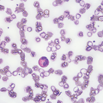
EGE can rarely cause eosinophilic ascites. Many case reports have shown EGE is associated with peripheral blood eosinophilia, as well as eosinophilic ascites. No diagnostic criteria have been proposed for eosinophilic ascites, and reports range from 23–95% eosinophils in ascites.6-9
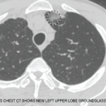
The ascites fluid analysis showed 28% eosinophils before the patient started steroid therapy. However, the stomach biopsy that showed 35 eosinophil/HPF was taken after the steroid regimen began for his RA flare. This could have shown a blunted eosinophil count in the tissue because he was already partially treated for his EGE with the prednisone prescribed for his RA. Likewise, although his CT scan showed dilated small bowel loops suggestive of an obstruction, he had no obstructive symptoms or abdominal pain, which may also reflect the fact that he was already being partially treated for his EGE.
The connection between RA and peripheral eosinophilia has been established. However, less is known about RA and eosinophilic infiltration of tissue, particularly in the GI tract. Some case reports have hinted at the connection between EGE and RA.10-14 Most patients showed improvement with steroid treatment (or an increase in steroid dosage if the patient was already on steroids).
Based on our literature search, there is a clear link between EGE and eosinophilic ascites, and a possible association between RA and EGE. To our knowledge, our case report is the first to link RA and EGE leading to eosinophilic ascites.


