“Maybe advanced imaging technology will help us to monitor and target treatment much better,” he said.
Imaging for Gout
In imaging for gout, Kay Hermann, MD, senior consultant in the department of radiology at Charite University Hospital in Berlin, cautioned that X-rays are not sensitive at the early stages.
In one study of 27 patients with gout, but with normal X-rays, MRI and ultrasound were performed on a symptomatic joint and an asymptomatic joint. Researchers found that a large percentage of patients with normal radiographs can have destructive arthropathy. Fifty-six percent of the patients were found to have erosions, with MRI much more sensitive than ultrasound.3
Dual-energy CT, with two X-ray beams used simultaneously, can be useful for specific detection of gouty tophi, while ultrasound is suited for finding faint gouty deposits on the surface of cartilage, Dr. Hermann said.
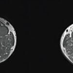
MRI, he said, can be used in more “unusual settings,” with the advantage that tophus volume measurements are possible.
Thomas Collins is a freelance medical writer based in Florida.
References
- Weber U, Lambert RG, Østergaard M, Hodler J, Pedersen SJ, Maksymowych WP. The diagnostic utility of magnetic resonance imaging in spondylarthritis: An international multicenter evaluation of one hundred eighty-seven subjects. Arthritis Rheum. 2010;62:3048-3058.
- Weckbach S, Schewe S, Michaely HJ, Steffinger D, Reiser MF, Glaser C. Whole-body MR imaging in psoriatic arthritis: Additional value for therapeutic decision making. European J Radiol. 77:149-155.
- Carter JD, Kedar RP, Anderson SR, et al. An analysis of MRI and ultrasound imaging in patients with gout who have normal plain radiographs. Rheumatology. 2009;48:1442-1446.

