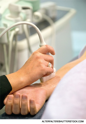
PARIS, FRANCE—Ultrasound can be more useful in rheumatic diseases than many rheumatologists realize, according to a panel of experts gathered at the Annual European Congress of Rheumatology (EULAR 2014) in June.
Each presenter answered a question about ultrasound in the setting of rheumatology—touching on joint counts in RA, obtaining biopsies and Sjögren’s syndrome.
Do we need a different ultrasound joint count for diagnosing & monitoring remission in rheumatoid arthritis?
Annamaria Iagnocco, MD, professor of rheumatology at the University of Rome, said that although ultrasound has been shown to be capable of predicting joint damage and short-term relapse in patients who are in remission—and many scoring models exist to make global assessments of these patients—there is no real consensus on how many joints need to be looked at in a global assessment or what joints those should be.
In fact, the scoring systems in the literature vary dramatically, with some including just one joint and others including up to 70.
Feasibility is an important concern, Dr. Iagnocco said. “Reduced joint count is particularly useful in terms of clinical practice for evaluating patients with rheumatoid arthritis while we are studying them in the assessment during our physical examination and clinical assessment,” she said.
The literature, when studies are compared side by side, reflects essentially an arbitrary approach to determining what joints to include.
In a study at Dr. Iagnocco’s center, a 12-joint reduced count approach was compared with a six-joint approach, which included the wrist, second metacarpophalangeal joint and knee on both sides. They found comparable results between the two, she said.1
“We found that this system was very quick to perform,” Dr. Iagnocco said. “When you have more than 12–20 joints to examine, this is very time consuming. But a global assessment is very important for diagnosis, for monitoring and for remission of the disease.”

What is the role of ultrasound in guided synovial biopsies?
Stephen Kelly, MD, consultant rheumatologist at Mile End Hospital in London, said that using ultrasound to help with getting samples of synovial tissue is a worthwhile approach.
Traditionally, samples have been obtained using arthroscopy, and patients have been referred to orthopedic surgeons. Some rheumatologists do it themselves, but it’s time consuming and expensive.
The samples are useful both for diagnosis and research—particularly in assessing whether the samples can be useful in predicting response to therapies, Dr. Kelly said.
In a study Dr. Kelly helped lead, researchers looked at the tolerability, safety and yield of synovial tissue in an early arthritis cohort using a minimally invasive, ultrasound-guided biopsy approach. In the procedure, a local anesthetic is injected into the skin and a small amount into the synovial space. Ultrasound is used to guide a semi-automated, guillotine-type needle.
A total of 93 consecutive biopsy procedures from 57 patients were assessed. The patients were recruited as part of the Pathobiology of Early Arthritis Cohort study. No significant complications were reported after the procedure. Nor were there any significant differences between before and after when it came to pain, swelling and stiffness.2
A median of 14 biopsy samples was obtained from each procedure. And 93% of the procedures yielded quality tissue.
When asked whether they would do it again, the vast majority of the patients said they were either very likely or somewhat likely to agree to it, Dr. Kelly said.
The yield of quality tissue is related to the degree of synovial thickening—with grade 3 giving an excellent yield. Overall, 92.5% of the samples were grade-able, and even those joints with minimal amounts of synovial thickening still had 45% graded biopsies, Dr. Kelly said.
He emphasized how important it is that the procedure be repeatable, so synovial tissue can be retrieved from the same joint at different times for research purposes. They also found that samples can be scanned and used later for comparison because they don’t show changes over time.
Dr. Kelly said a minimum of six samples will generally be representative of a large joint and four for a small joint. Getting that amount requires 8–10 samples, but it’s feasible to acquire 15–20 samples during a biopsy, allowing tissue to be processed for RNA and gene expression when required.
He said those considering the approach need to have a good basis in ultrasound training and need to be doing guided injections.
“I think this is a safe and well-tolerated procedure,” Dr. Kelly said. “Our patients are happy to have it performed repeatedly. You can access small and large joints, and get good quality tissue from histological grading and RNA. It’s reproducible and training certainly seems to be feasible.”
Researchers found that adding a Sjögren’s ultrasound score into the ACR criteria boosted sensitivity to 84%, compared with 64% with ACR alone.
How should ultrasound be applied in Sjögren’s syndrome?
Using ultrasound along with the American College of Rheumatology’s classification criteria for Sjögren’s improves diagnoses, said Sandrine Jousse-Joulin, MD, a rheumatologist in the Ultrasound Unit at La Cavale Branch Hospital in Brest, France.
In a study done earlier this year, researchers at the hospital studied 101 patients with suspected primary Sjögren’s syndrome. The cases in doubt were diagnosed by a blinded panel of experts. The patients had the four major salivary glands assessed by ultrasound.
Researchers found that adding a Sjögren’s ultrasound score into the ACR criteria boosted sensitivity to 84%, compared with 64% with ACR alone. Specificity dropped only slightly, falling from 91% to 89%.
Researchers at her center also wondered whether ultrasound might be able to replace minor salivary gland biopsy in the American-European Consensus Group criteria. They found they got better results when ultrasound assessment was added to the AECG criteria, with a sensitivity of 87% and a specificity of 96%. When ultrasound was substituted for the biopsy, sensitivity was just 68%, Dr. Jousse-Joulin said.
“US [ultrasound] could [be used to] follow primary Sjögren’s syndrome patients in evaluating structural abnormalities,” she said. “US has proved its importance to be added to the AECG criteria, but studies are required to evaluate this tool as an outcome measure.”
Thomas R. Collins is a freelance medical writer based in Florida.
References
- Perricone C, Ceccarelli F, Modesti M, et al. The 6-joint ultrasonographic assessment: A valid, sensitive-to-change and feasible method for evaluating joint inflammation in RA. Rheumatology (Oxford). 2012 May;51(5):866–873.
- Kelly S, Humby F, Filer A, et al. Ultrasound-guided synovial biopsy: A safe, well-tolerated and reliable technique for obtaining high-quality synovial tissue from both large and small joints in early arthritis patients. Ann Rheum Dis. 2013 Dec 13 [Epub ahead of print].
- Cornec D, Jousse-Joulin S, Marhadour T, et al. Salivary gland ultrasonography improves the diagnostic performance of the 2012 American College of Rheumatology classification criteria for Sjogren’s syndrome. Rheumatology (Oxford). 2014 April 4 [Epub ahead of print].

