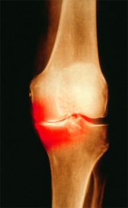
Colored X-ray of the knee of a patient with OA. Patients who test positive for anti-citrullinated protein antibodies show signs of bone erosion before they have clinical signs of arthritis.
Image Credit: Scott Camazine/sciencesource.com
ROME, Italy—The role of ACPA positivity in osteoarthritis is coming more into focus, with results from recent studies by researchers in Germany showing that ACPA positivity apparently works in concert with rheumatoid factor positivity to enhance bone erosion and that ACPA-positive patients show signs of bone erosion before they have clinical signs of arthritis.
The findings were presented at a session during EULAR 2015, the annual congress of the European League Against Rheumatism (EULAR).
Stephanie Finzel, MD, a resident in the rheumatology department at the University of Erlangen and a researcher on several recent studies on the topic, said these latest findings underline the importance of testing for anti-citrullinated protein antibodies (ACPA) positivity.
ACPAs & RF
In one study, the metacarpophalangeal joints (MCPJs) of 242 patients were scanned using high-resolution peripheral quantitative computed tomography (HR-pQCT). Then the number of erosions and their sizes were evaluated in 238 of them.1
A total of 112 patients showed positivity for both ACPAs and rheumatoid factor (RF), 28 were positive only for RF, 29 only for ACPAs and 29 for neither. The number of erosions and their sizes were highest in those both ACPA and RF positive, and the differences were significant in erosion number compared with those who were negative for both (p=0.001) and significant for erosion size compared with those who were ACPA negative (p<0.001).
They also found that RF influenced erosion number and size in patients who were ACPA positive, but not in ACPA-negative patients.
Researchers at the university also scanned the MCPJ of 10 people who were positive for ACPA, but with no signs of arthritis, with HR-pQCT. Ten healthy controls were also scanned, and the results for bone mineral density, cortical thickness and trabecularity were compared.2
The findings showed that it’s possible, with HR-pQCT, to measure bone changes in ACPA-positive patients without clinical signs of RA. Alterations were seen—mainly cortical thinning and fenestration in the cortical area.
Dr. Finzel said they support the idea that bone changes in RA are probably not due exclusively to inflammation and synovitis, but “occur in the phase of breakdown of immune tolerance” and ACPAs have the potential to be pathogenic.
“Bone in early rheumatoid arthritis likely undergoes vehement changes already in the very first weeks after the onset of swelling and pain,” she said. “Highly sensitive diagnostic tools, such as HR-pQCT, MRI or musculoskeletal ultrasound, are required to be able to diagnose correctly and treat early.”
Dr. Finzel also said the findings leave room to wonder whether it would be worthwhile to scan the general population during preventive check-ups.
Adaptive Immunity & Bone
In another line of research on the immune system and osteoarthritis, researchers at the University of Tokyo have shown in mice that the level of osteoclastogenesis is determined by the signaling strength of Fcy receptors. That signal strength is determined by a combination of factors, including the availability of their immune-complex ligands and the expression of activating and inhibiting receptors.3

Dr. Takayanagi
Autoantibody production and formation of the immune complex are frequently seen in autoimmune diseases causing bone loss. But these findings show that there is a direct link between adaptive immunity and bone, and suggest that the immune complex plays a regulatory role in bone resorption, said Hiroshi Takayanagi, MD, PhD, professor of immunology at the university.
Translational Process
Another osteoarthritis study—presented by Simon Mastbergen, PhD, assistant professor of tissue regeneration and rheumatology at University Medical Center in Utrecht, The Netherlands—showed for the first time the translational process at work after joint distraction in osteoarthritis (OA). The findings build on previous work by the group on cartilage repair after joint distraction in dog models.4
Four dogs received joint distraction surgery 10 weeks after OA induction, and four others served as osteoarthritis controls. Four weeks after the procedures, the animals were sacrificed, and knee tissues were harvested and quantitative polymerase chain reaction analyses were conducted for more than 64 regenerative markers.

Dr. Mastbergen
In the OA control group, typical OA markers, such as metalloproteinases, collagen and apoptosis markers, were up-regulated, demonstrating the OA disease cascade in effect. But joint distraction caused down-regulation of typical OA markers and maintenance of important matrix remodeling genes for regeneration, such as aggrecanase, Dr. Mastbergen said.
Researchers also found that genes from several pathways tied to OA were expressed differently in the two groups — such as TGF-beta, Wnt and Notch signaling.
“Distraction is a good candidate for knee OA treatment resulting in prolonged clinical and structural changes,” he said. “This study demonstrates distinctively that joint distraction initiates a transcriptional regulation of several important regenerative genes, indicating that a reset of joint homeostasis can lead to cartilage repair in OA.”
Thomas R. Collins is a freelance medical writer based in Florida.
References
- Hecht C, Englbrecht M, Rech J, et al. Additive effect of anti-citrullinated protein antibodies and rheumatoid factor on bone erosions in patients with RA. Ann Rheum Dis. 2014 Aug 12. [Epub ahead of print]
- Kleyer A, Finzel S, Rech J, et al. Bone loss before the clinical onset of rheumatoid arthritis in subjects with anticitrullinated protein antibodies. Ann Rheum Dis. 2014 May;73(5):854–860.
- Negishi-Koga T, Gober HJ, Sumiya E, et al. Immune complexes regulate bone metabolism through FcRγ signalling. Nat Commun. 2015 Mar 31;6:6637.
- Wiegant K, Intema F, van Roermund PM, et al. Evidence for cartilage repair by joint distraction in a canine model of osteoarthritis. Arthritis Rheumatol. 2015 Feb;67(2):465–474.

