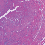
As noted above, the proposed model looks only at the probability of finding a positive biopsy, which is different than determining whether a patient has GCA. Many patients diagnosed with GCA actually have negative temporal artery biopsies, says Dr. Grayson.
Dr. Calabrese notes that there is nearly a complete absence of consideration in the model of the large group of GCA patients who have inflammatory presentations. These patients will likely, in the absence of head and neck symptoms, have negative biopsies and are now often diagnosed with vascular imaging.
It’s important to remember, according to Dr. Warrington, that the model authors are ophthalmologists and thus are focusing on patients who present with cranial symptoms such as headache, vision changes and jaw claudication. GCA, however, “comes in a lot of different shapes and sizes,” he says. “Some patients present with only fever of unknown origin; others have involvement of the aorta or subclavian artery and present with arm claudication; some present with stroke; some present with aortic aneurysm.”
Those presentations help a physician decide which diagnostic tool to use. For someone who presents with headache and vision loss, a temporal artery biopsy is probably the best tool. “But if someone has fever of unknown origin and no cranial symptoms, the likelihood of a positive biopsy is low,” Dr. Warrington says. “In that case we rely on imaging to make the diagnosis—CT, MRA or PET—to show inflammation of the large arteries.”
Recent clinical trials have revised inclusion criteria to include patients who were diagnosed by either biopsy or imaging. In some trials, up to half of patients are not diagnosed by biopsy but by imaging, he says.
The prediction model has some utility but should probably be applied only to patients with cranial symptoms and not patients with less common presentations. Incorporating temporal artery ultrasound for GCA diagnosis, as well as imaging large vessels (e.g., the chest arteries) into future models would be wise, Dr. Warrington says.
Regarding the model’s utility for rheumatologists, Dr. Grayson says he thinks the authors indicated its value in their conclusion. The researchers suggested, he says, “that perhaps their prediction model is most useful to help organize thinking but should not be a substitute for clinical decision making.”
Kathy Holliman, MEd, has been a medical writer and editor since 1997.
References
- Ing EB, Luna GL, Toren A, et al. Multivariable prediction model for suspected giant cell arteritis: Development and validation. Clin Ophthalmol. 2017 Nov 22;11:2031–2042.
- Ing EB, et al. The use of a nomogram to visually interpret a logistic regression prediction model for giant cell arteritis. Neuroophthalmology. Accepted for publication January 2018.
- Ing E, Pagnoux C, Tyndel F, et al. Lower ocular pulse amplitude with dynamic contour tonometry is associated with biopsy-proven giant cell arteritis. Can J Ophthalmol. 2017.
- Hunder GG, Bloch DA, Michel BA, et al. The American College of Rheumatology 1990 criteria for the classification of giant cell arteritis. Arthritis Rheum. 1990 Aug;33(8):1122–1128.

