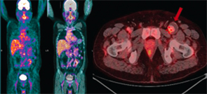
Figure 2. PET-CT showing low to moderate inflammatory activity with aneurismal dilation of the left common femoral artery with thrombus.
In 2014, a repeat PET-CT showed low to moderate inflammatory activity with aneurysmal dilation of the left common femoral artery (see Figure 2). He was advised to have surgery for the aneurysm after a period of increased immunosuppression. Six months later, he underwent left common femoral artery and superficial femoral artery aneurysm excision, with external iliac artery to superficial femoral artery interposition grafting (see Figures 3 and 4). Histologically, the aneurysmal segment showed disorganization of the elastic lamina of the media, with inadequate supportive fibrous tissue, and a large intraluminal thrombus (see Figure 5).
Four months following the surgery, he is stable and able to resume his normal activities.
Key points of this presentation:
- Although arterial narrowing is classical, aneurysms in TA are an important complication, and they can occur at multiple sites;
- Patients need regular monitoring, even if they are asymptomatic, because aneurysms can progress despite normal values of inflammatory markers and PET-CTs showing low disease activity;
- Rupture of these aneurysms can be catastrophic and, unlike atherosclerotic aneurysms, size may not be the only consideration for surgery;
- Timely surgical repair of aneurysms yields good results; and
- Patient education regarding the continuation of medication and regular follow-up are very important.
Discussion
Takayasu’s arteritis (TA) is a large-vessel arteritis commonly described in young Asians. It involves the aorta and its branches, with stenosis more commonly seen than aneurysms. In a large number of patients, the disease progresses without any symptoms until critical ischemia results in symptoms of claudication. Any part of the aorta can be involved, and the most commonly affected branches are the subclavian and common carotid arteries.1,2 Stenotic lesions are found in >90% of patients, whereas aneurysms are reported in approximately 25%.2 Stenotic lesions and aneurysms are associated with considerable morbidity and mortality. The consequences of TA are often severe, with 74% of patients reporting considerably compromised daily activities and 23% of patients unable to work.2
Disease monitoring is a great challenge to the treating physician because there are no markers, which can reliably predict disease activity. Inflammatory markers are notoriously fallacious in assessing disease activity. High-resolution ultrasound (US), magnetic resonance angiography (MRA), CT angiography (CTA) and PET have been used for diagnosis and monitoring disease activity (see Table 1). However, there still is no consensus on which imaging modality should be routinely used.


