Video: Knock on Wood| Webinar: ACR/CHEST ILD Guidelines in Practice
fa-facebookfa-linkedinfa-youtube-playfa-rss

An official publication of the ACR and the ARP serving rheumatologists and rheumatology professionals

  • Conditions
    • Axial Spondyloarthritis
    • Gout and Crystalline Arthritis
    • Myositis
    • Osteoarthritis and Bone Disorders
    • Pain Syndromes
    • Pediatric Conditions
    • Psoriatic Arthritis
    • Rheumatoid Arthritis
    • Sjögren’s Disease
    • Systemic Lupus Erythematosus
    • Systemic Sclerosis
    • Vasculitis
    • Other Rheumatic Conditions
  • FocusRheum
    • ANCA-Associated Vasculitis
    • Axial Spondyloarthritis
    • Gout
    • Lupus Nephritis
    • Psoriatic Arthritis
    • Rheumatoid Arthritis
    • Systemic Lupus Erythematosus
  • Guidance
    • Clinical Criteria/Guidelines
    • Ethics
    • Legal Updates
    • Legislation & Advocacy
    • Meeting Reports
      • ACR Convergence
      • Other ACR meetings
      • EULAR/Other
    • Research Rheum
  • Drug Updates
    • Analgesics
    • Biologics/DMARDs
  • Practice Support
    • Billing/Coding
    • EMRs
    • Facility
    • Insurance
    • QA/QI
    • Technology
    • Workforce
  • Opinion
    • Patient Perspective
    • Profiles
    • Rheuminations
      • Video
    • Speak Out Rheum
  • Career
    • ACR ExamRheum
    • Awards
    • Career Development
  • ACR
    • ACR Home
    • ACR Convergence
    • ACR Guidelines
    • Journals
      • ACR Open Rheumatology
      • Arthritis & Rheumatology
      • Arthritis Care & Research
    • From the College
    • Events/CME
    • President’s Perspective
  • Search

Fellow’s Forum Case Report: Diagnosing Antisynthetase Syndrome

Fawad Aslam, MBBS, and Elizabeth B. Russell, MD  |  Issue: May 2013  |  May 1, 2013

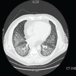
ILD is the most frequent manifestation, and in some cases the sole one, of antisynthetase syndrome.3 In about 20% of cases, the plain radiographs are read as normal, despite pulmonary involvement. When this diagnosis is being considered, high resolution CT scanning of the chest should be performed. Common findings include bilateral basal consolidations, diffuse patchy ground-glass opacities and subpleural prominence. On lung biopsy, NSIP is the most common pattern seen, although it is not specific for antisynthetase syndrome. Biopsy is important to rule out infections, granulomatous diseases, and vasculitis. It can also determine whether there is lung fibrosis. The absence of fibrosis predicts a better response to therapy. Although hemoptysis is not a regular feature of pulmonary involvement in antisynthetase syndrome, it has been reported.

Joint symptoms are more common in anti-Jo-1 positive patients and can mimic common inflammatory arthritides.4 RF, for example, can be positive in 10% to 20% of subjects with idiopathic pulmonary fibrosis (IPF). Patient with IPF can also have arthralgias, and it is important to look for secondary clues to an underlying connective tissue disease (CTD) before labeling any disease as idiopathic.

ad goes here:advert-1
ADVERTISEMENT
SCROLL TO CONTINUE

The lack of a unifying diagnosis, positive family history for a CTD, and absence of any identifiable environmental exposure or infection in the setting of a strongly positive SSA/SSB prompted the minor salivary gland biopsy, despite the fact that the patient did not have typical symptoms or the CT findings expected in SS. As noted, the biopsy was consistent with SS.

The utility of obtaining salivary gland biopsies in such settings can be debated, owing to false-positive rates and the significant variability in their interpretation. However, one study evaluating ILD patients without an underlying cause found 34% of patients having a biopsy suggestive of SS, of which 23% were negative for ANA, SSA, and SSB.5 About 23% of those with a positive biopsy denied sicca symptoms. The authors postulated that SS was subclinical in these patients. They also felt identification of a patient with an underlying disease, in this case SS—whether clinical or subclinical—conferred a better prognosis than the diagnosis of idiopathic ILD. It is difficult to say whether a salivary gland biopsy should be considered in an otherwise asymptomatic patient with an idiopathic ILD. SSA is frequently seen in association with anti-Jo-1, and evidence suggests an increased severity of ILD in patients with SSA positive antisynthetase syndrome.6

ad goes here:advert-2
ADVERTISEMENT
SCROLL TO CONTINUE

The lack of muscle symptoms in this patient contributed to the delay in arriving at the final diagnosis. It is important to note that myositis may not be one of the presenting features in antisynthetase syndrome. In one series of ILD patients with anti-Jo-1 antibodies, myositis was present in only 31% at initial presentation.3 The anti-Jo-1 presence can precede clinical myositis by years.

Page: 1 2 3 4 5 6 | Single Page
Share: 

Filed under:ConditionsOther Rheumatic Conditions Tagged with:Antisynthetase SyndromeInterstitial Lung Diseasepatient care

Related Articles

    What Rheumatologists Need to Know about Diagnosing and Managing Interstitial Lung Disease (ILD)

    December 1, 2012

    Patients with systemic sclerosis (SSc), poly-/dermatomyositis (PM/DM), or rheumatoid arthritis (RA) appear to carry the greatest risk for developing connective tissue disease-associated interstitial lung disease (CTD-ILD)
     

    How to Diagnose Antisynthetase Syndrome

    How to Diagnose Antisynthetase Syndrome

    March 17, 2017

    Antisynthetase syndrome (AS) is strongly associated with the presence of antibodies to aminoacyl-transfer RNA (tRNA) synthetases (ARSs) that are implicated in the pathogenesis of myositis and interstitial lung disease (ILD). Antibodies against eight antisynthetases have been identified and are detected in 16–26% of patients with idiopathic inflammatory myopathies (IIM).1 Serum assays for five of these…

    Scleroderma & ILD: Practical Tips on the Diagnosis & Management of Systemic Sclerosis-Associated Interstitial Lung Disease

    June 15, 2022

    No one-size-fits-all approach exists for the care and treatment of patients with systemic sclerosis (SSc) and SSc with pulmonary involvement. Here, experts discuss some best clinical practices for these patients.

    Fellow’s Forum Case Report: When Pulmonary Symptoms Point to Rheumatic Disease

    September 1, 2013

    A 48-year-old man with diffuse arthralgias and acute respiratory failure is diagnosed with antisynthetase syndrome

  • About Us
  • Meet the Editors
  • Issue Archives
  • Contribute
  • Advertise
  • Contact Us
fa-facebookfa-linkedinfa-youtube-playfa-rss
  • Copyright © 2025 by John Wiley & Sons, Inc. All rights reserved, including rights for text and data mining and training of artificial technologies or similar technologies. ISSN 1931-3268 (print). ISSN 1931-3209 (online).
  • DEI Statement
  • Privacy Policy
  • Terms of Use
  • Cookie Preferences