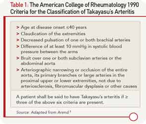
At age 28, she presented with hypertensive urgency and intestinal angina and was found to have renal artery stenosis. Magnetic resonance angiography (MRA) at that time revealed evidence of a large-vessel vasculitis of the abdominal aorta near the renal arteries, as well as the superior mesenteric artery, and she was diagnosed with Takayasu’s arteritis.
She was treated with high-dose glucocorticoids, azathioprine and infliximab infusions of 5 mg/kg every eight weeks. During tapering of her prednisone dose, she developed abdominal pain suggestive of intestinal ischemia. The dose and frequency of infliximab infusions were increased to 5 mg/kg every six weeks. On this regimen, she was able to taper off prednisone and then, four months prior to admission, to taper off azathioprine.
One month prior to admission she again developed postprandial abdominal pain, which improved after a short course of prednisone. The week prior to admission, the patient developed worsening headache and frothy urine, and she was found to have a blood pressure of 175/104 mmHg in the right arm at her infusion visit. She denied fevers, chills, weight loss, abdominal pain, arm or leg claudication, rash or other new symptoms.
On admission to the hospital, her blood pressure was 200/108 mmHg with no remarkable differences between all four extremities. ESR was 35 mm/hr, compared to a baseline of 7 mm/hr. Her complete blood count was unremarkable. Potassium was 5.1 mmol/L with a creatinine of 3.95 mg/dL. Urinalysis revealed an absence of red and white blood cells, and a urine protein to creatinine ratio was 0.47. Renin activity and aldosterone were both elevated at 52 pg/mL and 46.6 ng/dL respectively with a normal ratio, suggestive of renovascular disease. Duplex ultrasound was a limited study showing decreased flow in the right kidney, which appeared atrophic. Flow velocities were elevated in the left renal artery. An MRA without gadolinium revealed wall thickening of the abdominal aorta most prominent at the level of the superior mesenteric artery (SMA) extending to below the location of the renal arteries with wall thickening of the proximal SMA and left renal artery (see Figure 3). The right renal artery was diminutive with atrophy of the right kidney, similar to studies in 2011.


