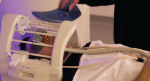
Mark Harmel / Science Source
Brain imaging can distinguish fibromyalgia patients from healthy controls with high sensitivity and specificity, according to two papers published nearly simultaneously in Pain late last summer, by groups at the Universities of Colorado and Michigan, respectively.
Somewhat surprisingly to the authors and others, in the Colorado study, which used both painful and nonpainful stimuli, the latter produced the strongest signals. If validated, the research may lead to a better understanding of fibromyalgia and improved treatments.1
“Both of these papers highlight the importance of sensory processing regions in experiencing chronic pain in fibromyalgia,” says Yvonne Lee, MD, MMSC, assistant professor at Harvard Medical School and Brigham and Women’s Hospital, who was not involved in the research. “Regulation of chronic pain may occur not only through classic pain pathways, but also via processes that affect sensitivity to a wide variety of stimuli.”
In the Colorado study, the first to be published, the investigators, including Tor Wager, PhD, and Jesus Pujol, MD, used fMRI to study the brain activity of 37 fibromyalgia patients and 35 healthy controls, as they were exposed simultaneously to various nonpainful visual, auditory and tactile clues (multisensory stimulation), and separately to painful pressure. Dr. Wager is professor of psychology and neuroscience, and director of the Cognitive and Affective Neuroscience Laboratory at his institution.
The multisensory stimulation included simultaneous exposure to flashing checker boards that alternated the positions of the white and black squares six times per second and auditory stimulation from each of 15 tones. At the same time, subjects were instructed to touch the tip of their right thumb to the fingers on that hand in rapid succession. “This approach allowed us to maximize signal power and challenge both sensory and motor systems efficiently,” the investigators write.
Nonpainful Stimuli Discriminate Better
The resulting data were analyzed using machine learning, a cutting-edge methodology that was applied to neuroimaging data to identify whether a person was a patient or a healthy control. Machine learning is a type of artificial intelligence that provides computers with the ability to learn without being explicitly programmed to do so. In this particular case, the investigators used a type of machine learning that Dr. Wager described as “a family of algorithms for finding patterns in complex data and using them to make accurate predictions.” It identified the patterns of brain activity with the biggest differences between patients and controls, distinguishing them with a 93% success rate.
These investigators also used fMRI to scan study subjects while they received painful pressure stimuli.
“The interesting thing to me is that the nonpainful stimuli were able to classify patients better than the painful stimuli,” says Richard Harris, PhD, associate professor, Chronic Pain and Fatigue Research Center, Anesthesiology, and associate professor, Division of Rheumatology, Internal Medicine, University of Michigan Medical School. “That implies to me that the problem in fibromyalgia is more of a sensory gain problem—like turning up the volume on your radio.” Thus, sensory amplification “may be the more relevant outcome to examine, the more relevant pathological factor driving the symptoms.”
“The complaints that fibromyalgia patients express about being annoyed by nonpainful sensory signals in daily life, such as noises—coming from the TV, for example—or sunlight, or even the contact of bed sheets, fit well with our results, showing strong abnormalities in the processing of these types of stimulus that are not normally painful, but that become unpleasant to patients,” says Marina Lopez-Sola, PhD, first author of the Colorado paper, who is a postdoctoral researcher at that university’s Cognitive and Affective Control laboratory.
All 3 Discomfort Sources Correlate
The Michigan study, which included 42 patients and 20 controls, also subjected patients separately to unpleasant but nonpainful (visual) stimulation, and to pressure pain on the thumbnail bed.2 The patients rated the levels of discomfort using the Gracely Box Scale, a scale ranging from 0 to 20. Patients also rated their current clinical pain. “For fibromyalgia, the pain is spontaneous,” explains Dr. Harris. “They have it all the time at different levels.”
The levels of all three sources of discomfort correlated closely in each patient. From that, “We concluded that visual stimulation is tapping into the neural mechanisms by which fibromyalgia patients experience pain,” says Dr. Harris. “This means that visual stimulation is tapping into some neurobiological structure that’s very intimately associated with the clinical pain the patient experiences, as well as the experimental pain,” he adds, referring to the insula, an area of the brain involved in pain perception.
On top of all that, patients treated with pregabalin who experienced pain relief from the fibromyalgia also showed a reduction in activation of the insula, says Dr. Harris. At the same time, they showed a reduced aversive response to light.
Although not definitive, the evidence from both studies is strong that fibromyalgia is caused within the central nervous system, rather than in the periphery, says Dr. Harris. “When you give people a drug that works to reduce pain and that negates or resolves the altered neurological activity, that suggests the brain is playing more of a causal role. And if that’s the case, that could point the way to potential treatments,” he says.
“We’d need a large clinical trial to prove that these changes we see are driving the pain,” says Dr. Harris. Such a trial would require scanning a group of subjects lacking chronic pain, to see which ones nonetheless had neural signatures of fibromyalgia. They would then have to be followed for at least two, and maybe as much as 10 years to see if significant numbers developed the pain. Then, if the findings showed that the pathology predated the pain, it would be possible to say that the neuroimaging results were not a consequence of the pain, he says.
The alternative hypothesis—supported by “some in the field,” says Dr. Harris, is that some chronic pain conditions— possibly including fibromyalgia—have a “peripheral generator.” Osteoarthritis inflammation or nerve damage in stroke—to name two of multiple possibilities—could send pain signals into the brain, he says. Those who favor the peripheral generator posit that the persistence of peripheral pain information coming into the central nervous system could cause the increase in pain, says Dr. Harris.
Benefits of the Research
The most immediate benefit for patients is that the imaging demonstrates the oft-denied reality of their symptoms, “providing them with an objective finding, a data point that says they have a real condition, which implies they can be treated,” says Dr. Harris.
If the research is validated, it likely will also become possible to use fMRI to determine which patients will respond to pain medication. Pain medications work on only about 30% of pain patients, including roughly that fraction of fibromyalgia patients, says Dr. Harris. The pattern of activity in the brain—which brain regions are active or inactive at the same time—may be able to predict treatment response, he says. That would save money and time for both patients and doctors.
Other benefits may accrue to patients with pains that are generally assumed to be specific to a body part, such as osteoarthritis in a knee or pelvic pain in women, rather than generalized, as in fibromyalgia. The problem is that sometimes excising the peripheral body part, such as with knee replacement or hysterectomy, occasionally fails to cure the pain, potentially because the pain is generated in the brain. And physicians frequently don’t take a thorough history regarding pain symptoms. “Very rarely do they assess how widespread the pain is,” says Dr. Harris. “Do they have pain in other areas? Do they have these other types of disorders that co‑aggregate with fibromyalgia?”
Although not presently possible, Dr. Harris says that in the future, “Imaging may be able to confirm these cases, and it could also discover new pathologies that are not currently captured with self report.”
“This is definitely worth investigating,” says Dr. Lee.
Ultimately, says Dr. Wager, the hope is that research, such as this, will lead to a thorough understanding of fibromyalgia. Presently, fibromyalgia is a syndrome with many subgroups, he says. Patients with ample discomfort related to touch, hearing or vision, but without specific pains in specific places, “may have some underlying problem, which may be related to inflammation or systemic autoimmune disease or broader kinds of brain psychopathology, more like generalized anxiety disorder or depression.” The goal, he says, is to identify brain features that can provide clues for defining subgroups that have more homogeneous neuropathology.”
“Currently, fibromyalgia diagnosis consists of individuals with similar symptoms but who have different pathways leading to those symptoms,” says Dr. Lee. “Thus, some of these methods may be helpful in identifying specific subtypes of patients with fibromyalgia, which in turn may enable better treatments targeted to specific subtypes.”
Potential treatment technologies—still well in the future for these applications—may include transcranial magnetic stimulation or transcranial direct current stimulation in the insula or other parts of the brain that may be found to be involved in fibromyalgia. “You may be able to inhibit specific brain areas without requiring surgery,” says Dr. Harris.
Says Dr. Wager, “We need to identify patients based on underlying pathology rather than just symptoms.”
David C. Holzman writes on medicine, science, environment and energy from Lexington, Mass.
References
- López-Solà M, Woo CW, Pujol J, et al. Towards a neurophysiological signature for fibromyalgia. Pain. [2016 Aug 31, Epub ahead of print] 2017 Jan;158(1):34–47.
- Harte SE, Ichesco E, Hampson JP, et al. Pharmacologic attenuation of cross-modal sensory augmentation within the chronic pain insula. Pain. 2016 Sep;157(9):1933–1945.
Another Anatomical Marker for Fibromyalgia
In earlier research, Richard Harris, PhD, of the University of Michigan Medical School, and his group previously posited that the activation of the insula is closely tied to a network called the Default Mode Network in fibromyalgia.1 This is a network of brain areas that are activated when a person is thinking introspectively, or “essentially assessing the status of their homeostasis,” says Dr. Harris.
Although the Default Mode Network is not directly activated by pain per se, it is more connected to areas of the brain that are involved in pain, notably the insula, in some pain conditions, such as fibromyalgia. “The insula is definitely an area that lights up in response to pain,” says Dr. Harris.
In previous studies, they showed that “the connectivity between the Default Mode Network and the insula correlates so well with the level of pain a patient feels that “it’s coming close to being a biomarker,” said Dr. Harris’s coauthor and University of Michigan colleague, Daniel J. Clauw, MD, referring to a different study by this group.2
References
- Napadow V, LaCount L, Park K, et al. Intrinsic brain connectivity in fibromyalgia is associated with chronic pain intensity. Arthritis Rheum. 2010 Aug;62(8):2545–2555.
- Loggia ML, Kim J, Gollub RL, et al. Default mode network connectivity encodes clinical pain: An arterial spin labeling study. Pain. 2013 Jan;154(1):24–33.