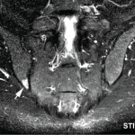
Objective: Low-grade bone marrow edema (BME) has been reported in the sacroiliac (SI) joints of 25% of healthy individuals and patients with nonspecific mechanical back pain, thus challenging the specificity and predictive value of magnetic resonance imaging (MRI) for the identification of early spondyloarthritis (SpA). It is unknown whether stress injury in competition sports may trigger BME. This study sought to explore the frequency and anatomic distribution of SI joint MRI lesions in recreational and elite athletes.
Methods: After pretest calibration, semicoronal MRI scans of the SI joints of 20 recreational runners before and after running and 22 elite ice-hockey players were assessed for BME and structural lesions. Three readers assessed the MRI scans in a blinded manner, using an SI joint quadrant–based module; scans from tumor necrosis factor–treated patients with SpA served for masking. The readers recorded subjects who met the Assessment of SpondyloArthritis international Society (ASAS) definition of active sacroiliitis. For descriptive analysis, the frequency of SI joint quadrants exhibiting BME/structural lesions, as concordantly recorded by at least two of the three readers, and their distribution in eight anatomic SI joint regions (the upper and lower ilium and sacrum, subdivided in anterior and posterior slices) were determined.
Results: The proportions of recreational runners and elite ice-hockey players fulfilling the ASAS definition of active sacroiliitis, as recorded concordantly by least two of the three readers, were 30–35% and 41%, respectively. In recreational runners before and after running, the mean ±SD number of SI joint quadrants showing BME was 3.1 ± 4.2 and 3.1 ± 4.5, respectively, while in elite ice-hockey players, it was 3.6 ± 3.0. The posterior lower ilium was the single most affected SI joint region, followed by the anterior upper sacrum. Erosion was virtually absent.
Study limitations include the small sample size and the lack of data from subjects with other conditions that may potentially confound the lesion signature on SI joint MRI, such as multiparity or heavy labor work.
Conclusion: In recreational and elite athletes, MRI revealed BME in an average of three or four SI joint quadrants, meeting the ASAS definition of active sacroiliitis in 30–41% of subjects. The posterior lower ilium was the single most affected SI joint region. These findings in athletes could help refine data-driven thresholds for defining sacroiliitis in early SpA.
Excerpted and adapted from:
Weber U, Jurik AG, Zejden A, et al. Frequency and anatomic distribution of magnetic resonance imaging features in the sacroiliac joints of young athletes: Exploring ‘background noise’ toward a data-driven definition of sacroiliitis in early spondyloarthritis. Arthritis Rheumatol. 2018 May;70(5):736–745.


