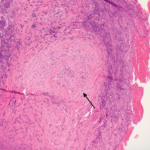
GPA was previously described as limited or generalized, with the latter comprising about 10% of cases. Limited GPA referred to all patients without renal involvement, often presenting with granulomas without vasculitis. More recently, this classification has become less popular, because limited disease could still be life threatening, such as in our patient’s case. Instead, it is useful to think of the disease as part of a spectrum—with localized disease that can progress to early systemic involvement, followed by generalized disease with imminent organ failure (organ threatening) and, eventually, severe disease that becomes life threatening. The clinical diagnosis should ideally be confirmed with a biopsy of the kidneys, skin or lungs (thoracoscopically). Transbronchial lung and nasal biopsies are typically unhelpful.3
Constitutional symptoms are often present, including fever, weight loss, fatigue, and malaise. Skin and musculoskeletal involvement commonly affects patients at some point, manifesting as palpable purpura, ulcers, nodules and arthralgia/myalgia usually in the absence of synovitis. Eye involvement is common and can sometimes be the initial presentation. Proptosis affects approximately 15% of patients, and there can be scleritis, episcleritis, uveitis and conjunctivitis.
Approximately half of all patients present with chronic sinusitis, with chronic inflammation present in the nasal mucosa. This can lead to purulent discharge, ulceration or granulomas of the mucosa, as well as septal perforation and nasal cartilage destruction. Stridor is the most worrisome sign of upper airway involvement.3
Approximately half of all patients present initially with pulmonary involvement, although many of these patients are asymptomatic despite positive chest X-ray findings. Renal disease affects about 15–30% of GPA patients on presentation—and eventually affects a majority. Very mild cases—such as in our patient—can present with minimal involvement, such as isolated hematuria and/or proteinuria, while others can have asymptomatic GN with proteinuria, pyuria and hematuria with casts. More severe cases can present with a pauci-immune, crescentic GN. Large vessel renal vasculitis and granulomas are extremely uncommon.4,5
How can we distinguish between the diseases in our differential diagnosis? In addition to the pulmonary involvement described thus far, the clinical entities described above are also known for their renal involvement. After an initial diagnostic workup, our patient’s differential was narrowed to the pulmonary-renal syndromes, a term describing the clinical entities that commonly cause diffuse alveolar hemorrhage and glomerulonephritis.
The three major causes are: 1) ANCA-associated vasculitides (AAV) (e.g., GPA, MPA, EGPA; 2) anti-glomerular basement antibody disease (e.g., GS); and 3) SLE.5 The final diagnosis relies on the specific pattern of clinical, serologic, pathologic and radiographic features. Although serologic testing can help, the turnaround time for some tests, such as ANCA, can be three to seven days, which makes it important to make a presumptive clinical diagnosis.


