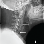
X-rays of her hands showed only soft tissue swelling, and an MRI of her C-spine showed joint effusion at the
C1–C2 level. Her left hip X-ray showed an irregular contour of the acetabular rim that, in the radiologist’s opinion, was a normal variant of the hip joint. During that time, although juvenile idiopathic arthritis (JIA) was suspected, in the absence of any erosive changes on imaging and due to very poor compliance, disease-modifying anti-rheumatic drugs (DMARDs) were deferred.
Additional test results included negative Lyme, normal uric acid and negative anti-cyclic citrullinated peptide (anti-CCP). Naprosyn liquid was advised, and her parents were asked to bring her back for follow-up in two months.
Over the course of the next three years, she continued to complain of pain of her right big toe, and in February 2016 she started complaining of pain over her right wrist. An X-ray of her wrist at that time showed erosive changes of the carpal bones, the ulnar styloid and the base of the metacarpals (see Figure 1) An MRI of the toe revealed erosive changes at the first metatarsophalangeal (MTP) joint.
Methotrexate was advised, but the mother declined, and instead hydroxychloroquine was started. Later, when the swelling and tenderness were only partially relieved, methotrexate was finally agreed upon, and the patient became completely pain free with 10 mg of methotrexate weekly.
On her most recent visit in December 2017, she still had residual swelling of the right big toe, but with no pain. Because of the erosive disease and persistent swelling, etanercept was suggested, but because the patient was asymptomatic it was refused.
Discussion
Down syndrome is the most common aneuploidy in humans and is considered the most common genetic cause of intellectual disability. It is the most frequent symptomatic aneuploidy compatible with survival and is strongly associated with maternal age. The underlying genetic cause of Down syndrome is the same in all those affected, but the clinical presentation varies.1
Several musculoskeletal abnormalities are associated with Down syndrome, including hypermobility of the joints, as well as low bone density, resulting in frequent fractures of both long bones and vertebral bodies in this population.2 The cause of low bone mass in Down syndrome, according to McKelvey et al., is attributable to low osteoblastic bone formation and inadequate bone accumulation, rather than to bone resorption.3 The study measured circulating biochemical markers—serum procollagen type-1 intact N-terminal propeptide (P1NP) to evaluate bone formation and serum C-terminal peptide of type-I collagen (CTx) to evaluate bone resorption—in adults with Down syndrome and compared them with control adults without Down syndrome.

