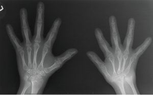
Figure 1: An X-ray of the hands, showing extensive erosion over the ulnar styloid, carpal bones and the carpometacarpal joints of the right side (A), along with abnormal shortening and thinning of diaphysis over the right second through fourth metacarpal bones (B). The X-ray of the left hand appears normal.
Down syndrome (trisomy 21) is one of the most common chromosomal abnormalities. According to the Genomic Resource Centre of the World Health Organization, each year 3,000–5,000 children are born with this chromosome disorder, and about 250,000 families have at least one member with Down syndrome in the U.S.
Down syndrome is caused by numerical aneuploidy, in which the child is born with an extra copy of chromosome 21. This chromosomal aberration affects various systems of the body, mainly neurological, cardiac, endocrinal, gastrointestinal and skeletal.
In this article, we discuss a case of trisomy 21 with a special emphasis on its effect on musculoskeletal health.
Case Presentation
A 9-year-old Caucasian female with Down syndrome was referred to the pediatric rheumatology clinic at Dartmouth in 2011, with complaints of bilateral lower extremity joint pain that had started about two years before. She had no associated history of fever, rashes or vision changes. On examination her joints were hypermobile but without active synovitis or tenderness. Her labs were negative (or normal) for rheumatoid factor (RF), erythrocyte sedimentation rate (ESR), anti-nuclear antibodies (ANA) and C-reactive protein (CRP), and she had a normal thyroid stimulating hormone (TSH) result. Her past medical history was significant for repaired congenital heart disease (bicuspid aortic valve with hypoplastic aortic arch and co-arctation of the aorta), hypothyroidism (controlled with thyroid hormone replacement therapy) and developmental delay. She had no significant family or social history. Because she had no other signs of inflammation, her joint pain was thought to be secondary to her hypermobility, and she was prescribed ibuprofen on as-needed basis and asked to follow up in three months.
Three years later, in June 2014—after missing many follow-up appointments—she came to the clinic again, this time with a six-month history of pain involving the left hip, left foot, cervical spine (C-spine) and interphalangeal joints of both hands, along with right big toe swelling secondary to an injury, but with no evidence of fracture.
On examination, she presented with swelling and tenderness over the interphalangeal joints of the hands and decreased flexion and extension of the C-spine. Again, she had a negative ANA, negative human leukocyte antigen (HLA) B27 and a negative RF. But her ESR was elevated at 23 mm/hr (normal: 0–20 mm/hr), and her CRP was elevated at 7.1 mg/L (normal: <3 mg/L).
X-rays of her hands showed only soft tissue swelling, and an MRI of her C-spine showed joint effusion at the
C1–C2 level. Her left hip X-ray showed an irregular contour of the acetabular rim that, in the radiologist’s opinion, was a normal variant of the hip joint. During that time, although juvenile idiopathic arthritis (JIA) was suspected, in the absence of any erosive changes on imaging and due to very poor compliance, disease-modifying anti-rheumatic drugs (DMARDs) were deferred.
Additional test results included negative Lyme, normal uric acid and negative anti-cyclic citrullinated peptide (anti-CCP). Naprosyn liquid was advised, and her parents were asked to bring her back for follow-up in two months.
Over the course of the next three years, she continued to complain of pain of her right big toe, and in February 2016 she started complaining of pain over her right wrist. An X-ray of her wrist at that time showed erosive changes of the carpal bones, the ulnar styloid and the base of the metacarpals (see Figure 1) An MRI of the toe revealed erosive changes at the first metatarsophalangeal (MTP) joint.
Methotrexate was advised, but the mother declined, and instead hydroxychloroquine was started. Later, when the swelling and tenderness were only partially relieved, methotrexate was finally agreed upon, and the patient became completely pain free with 10 mg of methotrexate weekly.
On her most recent visit in December 2017, she still had residual swelling of the right big toe, but with no pain. Because of the erosive disease and persistent swelling, etanercept was suggested, but because the patient was asymptomatic it was refused.
Discussion
Down syndrome is the most common aneuploidy in humans and is considered the most common genetic cause of intellectual disability. It is the most frequent symptomatic aneuploidy compatible with survival and is strongly associated with maternal age. The underlying genetic cause of Down syndrome is the same in all those affected, but the clinical presentation varies.1
Several musculoskeletal abnormalities are associated with Down syndrome, including hypermobility of the joints, as well as low bone density, resulting in frequent fractures of both long bones and vertebral bodies in this population.2 The cause of low bone mass in Down syndrome, according to McKelvey et al., is attributable to low osteoblastic bone formation and inadequate bone accumulation, rather than to bone resorption.3 The study measured circulating biochemical markers—serum procollagen type-1 intact N-terminal propeptide (P1NP) to evaluate bone formation and serum C-terminal peptide of type-I collagen (CTx) to evaluate bone resorption—in adults with Down syndrome and compared them with control adults without Down syndrome.
The study shows the mean P1NP level in adults with Down syndrome is lower than in controls, with no significant difference in their CTx levels, thus emphasizing a newer paradigm in the treatment of osteoporosis in Down syndrome focused on increasing the amount of bone formation in this population.
Several musculoskeletal abnormalities are associated with Down syndrome, including hypermobility of the joints, as well as low bone density, resulting in frequent fractures of both long bones & vertebral bodies in this population.
Hypermobility is a combined effect of muscle hypotonia and excessive ligamentous laxity. Patellofemoral instability is a common issue, with an estimated incidence of 10–20%, and is generally treated with non-operative modalities, but sometimes may require soft tissue and bony procedures in recurrent symptomatic cases.4 Spinal conditions, such as atlanto-axial or occipital-cervical joint instability with a reported incidence of 10–15% and scoliosis of about 7%, are also some commonly encountered in the Down syndrome population.5-7
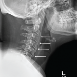
Reports have been made of significant osteoarthritic degenerative changes of the cervical spine with spur formation, narrowing of foramina, narrowing of the disc inner space and osteophyte formation on radiographic review of the C-spine of adults with Down syndrome between 26 and 42 years of age, emphasizing the importance of taking baseline radiographs of the C-spine in all Down syndrome children older than 5 years old and all Down syndrome adults older than 30.8
Foot abnormalities include pes-planus and first ray disorders, including metatarsus primus varus and hallux valgus, the treatment for which always begins with non-operative management, including orthotics or footware modification and surgical options when necessary.6,9,10
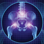
Down syndrome also affects the hips, causing slipped capital femoral epiphysis (SCFE), Perthes disease and hip instability.11 The etiology for SCFE in Down syndrome is unknown, although that for Perthes disease and hip instability is related to a multifactorial interplay of bony differences and soft tissue laxity with hypermobility.9,11 The treatment starts with immobilization and, depending on the severity and complexity, surgical interventions.
Arthritis in Down syndrome is less recognized, but as with other arthropathies, can result in chronic disability and functional impairment. The cause for this increased incidence of autoimmune phenomena is unknown. Cases of JIA with Down syndrome were described by Harbir Juj et al., in the Journal of Pediatrics, which stated the prevalence of arthropathy is 8.7/1,000—more than six times higher than JIA in general population.12
Harbir Juj et al. described nine children who had Down syndrome with JIA and a variety of presentations. All responded to different forms of anti-inflammatory medications to some extent. The researchers emphasized that, due to lack of awareness about the occurrence of inflammatory arthritis in Down syndrome, many children had to undergo unnecessary procedures or experienced delayed diagnosis, which led to a few cases of irreversible joint damage. Therefore, it’s very important to address early on any signs of inflammation in a population already at significant risk, to reduce their morbidity burden.
Our case illustrates the potential severity of this condition when not addressed in a conventional manner with timely introduction of DMARD therapy and, if necessary, biologic agents.
Conclusion
Down syndrome is a common genetic disease, affecting various systems of the body, including the musculoskeletal system. It mainly causes low bone density, joint instability and the less-documented arthropathy. The presentation of these pathologies might differ between cases, but with early suspicion and specific diagnostic tests, patients can have significantly less debilitating conditions. The purpose of reporting these children is to increase awareness of the association between trisomy 21 and arthropathies, along with other musculoskeletal disorders. Further investigation of this association may give clues to the relationship between genetic and immunologic factors in the pathogenesis of joint inflammation along with other musculoskeletal abnormalities and is highly encouraged.
 Prasanna Bastola, MBBS, is research fellow at Dartmouth Hitchcock Medical Center in the Department of Pediatric Rheumatology. She obtained her MBBS degree from Manipal College of Medical Sciences, Nepal, and is aspiring to become a pediatrician in the U.S.
 Daniel A. Albert, MD, is a rheumatologist at Dartmouth Hitchcock Medical Center and a professor of medicine and pediatrics at Geisel School of Medicine at Dartmouth and the Dartmouth Institute. Dr. Albert sees both adults and children with rheumatic disease, and his main areas of focus are RA and vasculitis.
References
- Antonarakis SE, Lyle R, Dermitzakis ET, et al. Chromosome 21 and Down syndrome: From genomics to pathophysiology. Nat Rev Genet. 2004 Oct;5(10):725–738.
- Van Allen MI, Fung J, Jurenka SB. Health care concerns and guidelines for adults with Down syndrome. Am J Med Genet. 1999 Jun 25;89(2):100–110.
- McKelvey KD, Fowler TW, Akel NS, et al. Low bone turnover and low bone density in a cohort of adults with Down syndrome. Osteoporos Int. 2013 Apr;24(4):1333–1338.
- Mendez AA, Keret D, MacEwen GD. Treatment of patellofemoral instability in Down’s syndrome. Clin Orthop Relat Res. 1988 Sep;(234):148–158.
- Ali FE, Al-Bustan MA, Al-Busairi WA, et al. Cervical spine abnormalities associated with Down syndrome. Int Orthop. 2006 Aug;30(4):284–289.
- Morad M, Kandel I, Merrick-Kenig E, Merrick J. Persons with Down syndrome in residential care in Israel: Trends for 1998–2006. Int J Adolesc Med Health. 2009 Jan–Mar;21(1):131–134.
- Pueschel SM, Scola FH, Tupper TB, Pezzullo JC. Skeletal anomalies of the upper cervical spine in children with Down syndrome. J Pediatr Orthop. 1990 Sep–Oct;10(5):607–611.
- Dyke DC Van, Gahagan CA. Down syndrome: Cervical spine abnormalities and problems. Clin Pediatr (Phila). 1988 Sep;27(9):415–418.
- Caird MS, Wills BPD, Dormans JP. Down syndrome in children: The role of the orthopaedic surgeon. J Am Acad Orthop Surg. 2006 Oct;14(11):610–619.
- Diamond LS, Lynne D, Sigman B. Orthopedic disorders in patients with Down’s syndrome. Orthop Clin North Am. 1981 Jan;12(1):57–71.
- Zywiel MG, Mont MA, Callaghan JJ, et al. Surgical challenges and clinical outcomes of total hip replacement in patients with Down’s syndrome. Bone Joint J. 2013 Nov;95-B(11 Suppl A):41–45.
- Juj H, Emery H. The arthropathy of Down syndrome: An underdiagnosed and under-recognized condition. J Pediatr. 2009 Feb;154(2):234–238.

