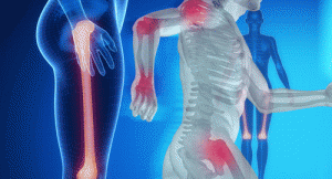
 The field of osteoimmunology has emerged to characterize what appears to be clinically important crosstalk between bone and the immune system. Although still in its infancy, the field has revealed that CD4+ T cells play a critical role in the communication between bone and the immune system. That said, investigators are still actively researching to what extent CD4+ regulatory T cells and memory T cells are able to influence bone cells. Given the murkiness of the current understanding, multiple investigators have focused their efforts on understanding whether or not CD4+ T cells can modulate the differentiation of bone-resorbing osteoclasts. The studies have examined both the crosstalk that occurs under normal physiological conditions, as well as the crosstalk that occurs during pathological conditions. Abdelilah Wakkach, PhD, a researcher at the University of Nice Sophia Antipolis in France and colleagues reviewed these osteoimmune interactions in the December 2015 issue of Frontiers in Immunology.1
Normal Physiological Conditions
Bone remodeling is highly regulated and relies on bone-forming osteoblasts (OBLs) and bone-resorbing osteoclasts (OCLs). Additionally, the bone marrow is considered the central hematopoietic organ in the body. Many cells appear to play an important role in the formation of the hematopoietic stem cell (HSC) niche in the bone marrow. These cells include OCLs, immature and perivascular mesenchymal cells, osteoprogenitors and OBLs. Specifically, mouse models of osteopetrosis suggest that the formation of the HSC niche is dependent on the ability of OSCs to resorb bone. After the HSC niche is formed, the bone undergoes endochondral ossification followed by a shift to hematopoiesis right before birth. At that point, hematopoietic progenitor cells hone to the HSC niche and cause it to expand. Both OCLs and macrophages appear to play important roles in regulating the HSCs outside of the bone marrow and recruiting them to the HSC niche.
After the system has stabilized, T cells represent approximately 3–8% of the total nucleated cells in the bone marrow. These T cells come from the blood to the bone marrow and can move from there back to the blood to migrate to other lymphoid organs. The bone marrow also serves as a reservoir for memory T cells, 25% of which are Foxp3+ regulatory T cells. Tissue-resident memory T cells are also maintained in the bone marrow, where they adapt to the local environment and ignore signals to migrate outside the bone marrow.
Pathology
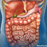
Researchers have long noted an association between chronic inflammation and bone destruction. This observation has led some to question whether immune cells play a role in bone remodeling. This question has been investigated in inflammatory bowel disease (IBD), which can take the form of either Crohn’s disease or ulcerative colitis. These diseases are often associated with extra-intestinal manifestations, in particular, bone loss. Patients with IBD have inflamed gastrointestinal mucosa that contains massive infiltrations of Th17 cells.
A mouse model of colitis has revealed that a large number of CD4+CD44+CD62L- memory T cells reside in bone marrow. This CD4+ T cell population appears to act as an osteoclastogenic population during IBD. Research is now focused on better characterizing these putative osteoclastogenic T cells. Studies in IBD suggest that they consist of TNF-α producing Th17 cells that are capable only of limited production of interferon ɣ. However, researchers have yet to identify how these cells are maintained in bone marrow.
Lara C. Pullen, PhD, is a medical writer based in the Chicago area.
Reference
- Wakkach A, Rouleau M, Blin-Wakkach C. Osteoimmune interactions in inflammatory bowel disease: Central role of bone marrow Th17 TNFα cells in osteoclastogenesis. Front Immunol. 2015 Dec 18;6:640. doi: 10.3389/fimmu.2015.00640. eCollection 2015.

