Lightspring / shutterstock.com
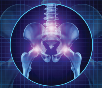
CHICAGO—Two experts presented insights on the diagnosis and treatment of low back and hip pain, including a refresher course on the mechanical structures involved, in Anatomy in a Day: Demystifying Low Back Pain and Lateral Hip Pain: New Patho-Anatomical Perspectives, a session at the 2018 ACR/ARHP Annual Meeting.
Low Back Pain
Avoid using such terms as non-specific low back pain, mechanical low back pain or idiopathic low back pain, said Jean H. Gillies, MD, clinical instructor of rheumatology at the University of British Columbia, Vancouver, Canada. Rheumatologists must identify specific causes of low back pain and offer their patients more effective therapies than just self-management.
Low back pain occurs in the region of the posterior trunk between the 12th rib and the gluteal folds. Hip pain may be located in the buttocks, the greater trochanteric region, groin or anterior, anterolateral or anteromedial thigh, according to Dr. Gillies.
“Why should rheumatologists be interested in low back pain?” Dr. Gillies asked. “Because low back pain has become a major global challenge, and the No. 1 cause of disability worldwide,” and was the subject of a major report in 2018 that included a call for action on better treatments for low back pain.1,2 According to the report, only a small fraction of patients with low back pain have a well-understood pathological cause, such as a vertebral fracture, so most cases are given the catch-all “non-specific” label.
“We’ve had no major advances in the diagnosis and treatment of low back pain over the past 50 years,” said Dr. Gillies. Most recommendations involve patient self-management with exercise or psychological support, which are ineffective.
The causes of back pain may fall into three clinical subsets: referred pain, lumbar spine disorders and radicular pain, said Dr. Gillies.3 Rheumatologists first screen patients with low back pain for red flags, including fracture, malignancy or spondyloarthropathies, which account for a small percentage of cases.4
In an Australian study of 1,172 low back pain patients, less than 1% had a specific, identified cause for their pain, and this gap is unacceptable, Dr. Gillies said.5
In her rheumatology practice, Dr. Gillies treats professional and Olympic athletes in many sports—as well as active weekend warriors—for back pain, but “if I followed the current international treatment guidelines, they wouldn’t help me at all, and it would mean [patients] don’t return to their sport,” she said.
Current guidelines include recommendations for low back pain ranging from heat to acupuncture to mindfulness-based stress reduction.6
Soft-Tissue Sources
Most low back pain cases involve a soft-tissue source, including mechanical enthesitis, such as ligament sprains, tendinitis or fasciitis, and bursitis and cutaneous nerve entrapments. These conditions may be acute or chronic. Sprains, tenderness or injuries to soft tissues of the entheses are common culprits for low back pain, including three in particular: iliolumbar ligament/thoraco-lumbar fascia (TLF) complex enthesopathy, sacrotuberous ligament/TLF complex enthesopathy and paraspinous process enthesopathy, involving the fascial attachments of the multifidus, erector spinae and the thoracolumbar
fascia, Dr. Gillies said.
Using diagrams, Dr. Gillies explained how to examine tender, soft-tissue sources of a patient’s low back pain: With the patient lying on their abdomen, palpate and mark the top of the iliac crests, followed by the inferior border and lateral border of the posterior superior iliac spine (PSIS). Then, palpate and mark the outline and border between the ilium and sacrum, and down the lateral border of the sacrum.
“These are the really common sites. When you go back to your office and someone has low back pain, have them lie face down and then press on these places: the medial iliac crest, the ilio-lumbar ligament, the side of the PSIS, and the paraspinous process entheses at S1 or S2.” Most patients react strongly to pressure at the source of their tenderness or pain, she said.
A specific source of back pain may be established in most cases, often due to mechanical enthesitis, said Dr. Gillies. Her opinion was influenced by a 1991 study showing 60% of patients who presented to an outpatient rheumatology clinic in the U.K. with low back pain had tenderness on palpation at the medial end of their iliac crests.7
Later, she treated a 48-year-old man with a 24-year history of low back pain and sciatica who presented with intermittent right buttock and right posterior thigh pain. “He had no constant pain, which is always good. I always say, if pain is intermittent, this should be treatable, because at times, it doesn’t hurt,” she said.
After palpation, she found his pain was not at the medial end of the iliac crest, but was very localized in an area the size of a can of tuna, and exacerbated by lifting. His back pain stemmed from an injury he sustained 25 years earlier while cross-country running.
He minimized his lifting activities in his veterinary work to manage his on-and-off pain. “He didn’t have any morning stiffness, but his maximum driving time was an hour, and then he’d have to stop and get out. He was constantly shifting when standing or sitting. Walking for him was unlimited and helped.”
Her patient had exquisite tenderness on the lateral crest of his PSIS. Dr. Gillies injected 10 cc of bupivacaine 0.25% into the ilial fibers of the proximal attachment of the right sacrotuberous ligament to confirm the site of his pain. “Bupivacaine lasts about five hours. If they’re pain free when they leave, and the pain comes back after exactly that amount of time, it’s an obvious diagnosis. But don’t tell them how long the medication lasts,” she said.
Her patient had no pain on his hour-long drive home, but his pain returned five hours later, as she expected. She then treated him with four injections every two weeks of 2 cc of triamcinolone (20 mg) and 3 cc of bupivacaine (0.25%), which eliminated his pain over the next year-and-a-half—except for one self-limiting episode after lifting a heavy box.
Although back pain from cutaneous nerve entrapment is less common, perineural injection treatment (PIT), or neural prolotherapy, of buffered dextrose 5% (D5W) around the swollen cutaneous nerves may be performed in the office to diagnose this cause within just 10 minutes, and it has no adverse effects—an approach studied in a 2016 case series of patients with medial superior cluneal nerve entrapment, said Dr. Gillies.8 A sports medicine specialist in New Zealand, John Lyftogt, MD, has used neural prolotherapy to rapidly relieve pain and restore mobility in low back pain patients, she said.9
‘One of the advantages of ultrasound is that you can characterize tendon, muscle & cortical bone abnormalities.’ —Ingrid Möller, MD, PhD
Sacroiliac Joint Pain
In a 2017 study of 100 patients with sacroiliac joint (SIJ) pain diagnosed by periarticular injection, researchers mapped the locations of patients’ pain, numbness and tingling.10 They found that 94 patients reported pain at or around the PSIS, and about 60% of patients reported lower limb pain and numbness or tingling sensations. Pain was detected mainly in the back, buttocks, groin and thighs, and numbness/tingling was mainly detected in the lateral and posterior thigh and posterior calf.
“How does the SI joint refer pain to the back?” asked Dr. Gillies. “It’s a misnomer. The low back goes from the 12th ribs to the gluteal folds, so you could divide it into two,” the lumbar region above the iliac crests and the buttock region below. “I’d suggest that when you talk about where the pain is, you say it’s in the lumbar region or it’s in the buttock region.”

BlurryMe / shutterstock.com
Structures in the buttock region do not refer pain to the lower back, because they are part of the lower back.11
“There are some anatomical concepts that really help you understand what happens in the sacroiliac joint. The synovial joints do three things: they roll, they slide and they spin. That’s it. But they can’t roll or slide on their own. These movements are coupled. They can spin on their own, but the rest of the movements are done together. That’s how joints move,” Dr. Gillies said. Codman’s paradox, a rotation of the shoulder joint, is an example of a coupled roll-and-slide movement. 12
“People always talk about sacroiliac joint subluxation,” such as chiropractic manipulation of the joint to treat irregular leg lengths, Dr. Gillies said. “If you dislocate a joint, the two joint surfaces are far apart. If you sublux them, that’s partial dislocation. I prefer to call this discentriclocation. When you extend your knee, there is a slide. It should end up resting in that central position. SI joints are not subluxed.” Patients with knee osteoarthritis often have uncoupled movements in this joint, she said.
Form closure and force closure work together in a stable, normal SI joint, and patients with malalignment of the SI joint often say they feel unstable, she said. Patients with SIJ pain may benefit from rolotherapy, injection of an irritant to cause localized inflammation that stimulates fibroblast hyperplasia and increased collagen production to promote healing.13
Another problem in low back pain are close-packed or loose-packed positioning of joints, said Dr. Gillies. While on a family vacation, her brother asked, “How do flamingos balance on one leg?” She explained that the joints of the bird’s standing leg are close packed. “Every joint has a close-packed position where it is like a pillar. In the flamingo, it’s in the ankle. So they can sleep standing on one leg with virtually no muscle energy whatsoever.”
How do birds achieve trunk stability to fly? “Flying birds have a fused cylinder, like an airplane. The only mobile bits are the neck and the distal part of the tail,” she said. “But how can we do that? We have to get that close-packed position in humans, where it is essentially fused, so when you bend forward, the sacrum moves. It’s called nutation.” A critical close-packed position in humans is full nutation in the L5 sacral joint, Dr. Gillies said.
To improve trunk stability for efficient load transfer, recommend that patients do alternating core exercises to strengthen abdominal and trunk muscles, although experts don’t fully agree on which exercises to include in a comprehensive program, Dr. Gillies concluded.14
Hip Pain Syndromes
To understand hip anatomy, look at the connections in the hip’s design, said Ingrid Möller, MD, PhD, assistant professor of anatomy and a rheumatologist at the University of Barcelona, Spain.
“The os coxae, or hip bone joints, are formed by the fusion of the ilium, ischium and pubis,” she said. “In this design, we want to confer elasticity to the pelvic ring and contribute to the transfer of weight from the spine to the lower limbs, as well as having a shock absorbing function that protects the spine. In that region, we have 27 muscles plus anatomical variations that cross the hip joint, acting as primary movers and dynamic stabilizers of the hip and the lower extremities.”
Hip anatomy adapted over time as the human gait evolved from quadrupedal to bipedal, said Dr. Möller. Our two-legged walk is facilitated by evolutionary adaptations of the lumbar spine, a shorter pelvis, double extension of the spine and hip, a longer femur, a larger ischium, and adaptations of muscle function in the hip region.15 These anatomical changes are relevant to understanding conditions like gluteal tendinopathy, the most prevalent tendinopathy in the lower extremities.16
Abductor and pelvic stabilizer muscles around the acetabulum “are really complex muscles, because the function changes from the anterior part of the joint to the medial and posterior aspects,” she said. They include the adductor muscles, lateral rotator muscles, and gluteal muscles, the gluteus minimus, medius and maximus.
“The gluteus minimus has in this lateral portion a function of flexion and medial rotation. But the gluteus medius makes more abduction. The gluteus maximus is doing extension and lateral rotation, plus abduction again,” Dr. Möller said. The goal of these multifunctional muscles is stabilization while we walk. “The position of the different muscles in relation to the acetabulum influences the action that they perform.”
Greater Trochanteric Pain Syndrome
Trochanteritis and bursitis of the hip are actually gluteal tendinopathy affecting the gluteus medius and minimus, and this is the primary pathology underlying greater trochanteric pain syndrome (GTPS), Dr. Möller said. Patients with GTPS present with “intermittent or chronic lateral hip pain with some radiation to the thigh or buttock area, and their pain is aggravated by some activities or lying on the affected side. But we lack validated or specific diagnostic criteria.”
GTPS has a multifactorial etiology that may involve biomechanics, abnormality of the lower limbs or abnormal force vectors across the hip joint, she said. It has a large prevalence, affects more females than males, and more often occurs after age 40.17 Dr. Möller shared a photo of her 60-year-old sister, a champion long-distance runner, to point out that GTPS prevalence is expected to rise as people live longer and more women engage in long-distance running.18
To examine patients with possible GTPS, try to reproduce the pain occurring with movement of the greater trochanter, said Dr. Möller. She suggested asking the patient to mimic the single leg stance of a flamingo. Differentiate GTPS from hip osteoarthritis by testing how well the patient can easily manipulate putting on shoes and socks, and with the flexion abduction external rotation (FABER) test to locate the source of the pain.19 Differentiate from low back pain by testing how well the patient rises from a seated position. If a patient has damage in the gluteus medius tendon, rising from a chair will be very difficult, she said.
“However, there is a weak association between physical examination and diagnosis, and if you trust in MRI [magnetic resonance imaging], a gold standard for many different conditions, there is no way to distinguish between asymptomatic and symptomatic patients,” Dr. Möller said.20 The hip and pelvic rotator mechanism involves synergy of two muscle groups: the trochanteric abductor muscles, including the gluteus minimus and medius, and the iliotibial tract tensor muscles, including the gluteus maximus, tensor fasciae latae and vastus lateralis, which may be clearly seen on an ultrasound scan.
Ultrasound’s Advantages
“One of the advantages of ultrasound is that you can characterize tendon, muscle and cortical bone abnormalities. We can see, better than with MRI, any calcifications of gluteal tendons, see excess fluid in the bursa, and assess, through sonopalpation, tenderness. We can do guided injection. And there is low cost, and it is available in many rheumatology departments,” Dr. Möller said. Ultrasound shows moderate sensitivity in prediction of surgically confirmed gluteus medius tears.21 “The limitations of ultrasound to detect tendon changes is mostly seen in obese patients.”
In the lower back, the gluteus maximus and contralateral latissimus dorsi are functionally coupled, assisting with trunk rotation, and stabilizing the lower lumbar spine and SI joints.22 Dr. Möller asked everyone to stand up and rotate their trunks, “and you will feel how your muscles are linked. This is the synergistic action of all these different muscles.” The gluteus maximus exerts compressive force at the SI joints by assisting load transfer between the lower extremities and the trunk, she said.23
“If you look at the upper, mostly abductor part of the gluteus maximus, you can see the insertion in the thoracolumbar fascia,” said Dr. Möller. “I’m a fan of the fascia. I can recognize their value. The thoracolumbar fascia is the most pain-sensitive deep tissue we have in the lower back.”24 The lower gluteus maximus is an extensor, and originates from the sacrum and sacrotuberous ligament. “We have two hip extensor mechanisms, one for speed and the other for power. The hamstrings provide rapid hip extension, whereas the gluteus maximus provides a powerful hip extension.”
Injury to the inferior gluteal nerve in this anatomical region causes difficulty in climbing stairs, standing up from a chair or walking. However, the hamstrings compensate in the standing position.
“This also helps us explain the extraspinal etiology of sciatic pain, or pain from the huge sciatic nerve, which will be aggravated by sitting, and you will have a tender point immediately lateral to the right ischial tuberosity,” said Dr. Möller.25 The deep, very powerful gluteus medius balances the side-to-side movement of the trunk during bipedal gait, and this muscle is a hip stabilizer and pelvic rotator.26 Every portion of the muscle contracts in a different way through each phase of gait.
“The superior gluteal nerve that innervates the gluteus medius is one nerve with three different branches for the three different portions of the muscle. That means you can damage one portion and you lose one of the activities during gait,” she said. “The posterior fibers of the gluteus medius are stabilizers, because they bring the femoral head closer to the acetabulum. The middle fibers, which are parallel to the axis of the femur, provide an important contribution in an extended posture, because abduction contributes to lateral stability during locomotion. The anterior fibers perform a function during internal rotation. The gluteus medius, this huge muscle, is powerful, and must perform the force of twice the body weight during a monopodial stance at the normal gait.”27 Fatty degeneration of the gluteus medius plays a role in loss of stability in the pelvis and may increase risk of falls.28 If a patient has a gluteus medius tear on one side, the contralateral side will drop when they put their weight on the affected hip when standing, she said.
Ultrasound can show hip abductor tears and pathological changes in the hip, such as calcifications, loss of fibrillar pattern, changes in tendon morphology, bone irregularities, and even the absence, or full tear, of the trochanteric tendon, said Dr. Möller. Conservative treatment of chronic hip abductor tear includes non-steroidal anti-inflammatory drugs (NSAIDs), exercises and local injections, but patients with persistent pain and functional limitation may need surgery.29
Progression with Aging
In the deep part of the trochanteric hip is the gluteus minimus, and here, tendon pathology and muscle atrophy increase with age, correlating with the progression of tendinosis to tendon tears, she said.30 This muscle shows fatty degeneration earlier than the gluteus medius, and atrophy on the anterior part of the muscle seen on an MRI can also predict falls in older adults.31 After total hip arthroplasty, some patients will experience greater trochanteric pain syndrome due to atrophy in the gluteus medius and minimus.32
Bursae are located over the lateral, posterosuperior and anterior facets of the hip, including the trochanteric or gluteofemoral bursa, which is often implicated in painful trochanteric bursitis. It is located caudal to the greater trochanter, beneath the iliotibial tract, where the gluteus maximus’ tendinous fibers are inserted, and it separates the iliotibial tract from the vastus lateralis.
“Bursitis can also be associated with the inflammatory pathology of the hip. That means in a patient with inflammation, you can have bursitis,” said Dr. Möller, who showed ultrasound images of an inflamed hip joint with distension of the hip capsule tissues and bursitis next to the gluteus minimus. Ultrasound-guided, local injection of corticosteroid into the affected hip “will produce an initial reduction in pain with 72–75% reported positive response at four weeks.”33
The iliotibial tract (ITT) is a longitudinal, fibrous band created by opposing forces pulling on the fascia lata, the deep fascia investing the muscles of the hip and thigh, she said. Proximal ITT syndrome includes degenerative tears of this fibrous band, which can occur in middle-aged to elderly women even when there has been no trauma. Proximal ITT syndrome may also be an overuse injury that causes pain and tenderness at the iliac tubercle.34 The ITT may compress the tendons at the gluteus medius and minimus at their trochanteric insertion in the excessive hip adduction position or at high ranges of hip flexion, causing clinical symptoms like lateral hip pain with prolonged sitting and difficulty rising from a chair.35
A thickened posterior ITT band moving over the external greater trochanter can be one of many causes of external snapping hip syndrome, Dr. Möller said. Contraction of the huge gluteus maximus may transmit force to the ITT and the fascia lata, and this pathology may contribute to distal ITT syndrome.36 Prolonged sitting may result in shortening of the tensor of the fascia latae, which may produce an anterior tilt of the pelvis and/or a medial rotation of the femur.
Susan Bernstein is a freelance medical journalist based in Atlanta.
References
- Hoy D, March L, Brooks P, et al. The global burden of low back pain: Estimates from the Global Burden of Disease 2010 study. Ann Rheum Dis. 2014 Jun;73(6):968–974.
- Clark S, Horton R. Low back pain: A major global challenge. Lancet. 2018 Jun 9;391(10137):2302.
- Maher C, Underwood M, Buchbinder R. Non-specific low back pain. Lancet. 2017 Feb 18;389(10070):736–747.
- Deyo RA, Weinstein JN. Low back pain. N Engl J Med. 2001 Feb 1;344:363–370.
- Henschke N, Maher CG, Refshauge KM, et al. Prevalence of and screening for serious spinal pathology in patients presenting to primary care settings with acute low back pain. Arthritis Rheum. 2009 Oct; 60(10):3072–3080.
- Qaseem A, Wilt TJ, MacLean RM, et al. Noninvasive treatments for acute, subacute and chronic low back pain: A clinical practice guideline from the American College of Physicians. Ann Intern Med. 2017 Apr 4;166(7):514–530.
- Collee G, Dijkmans BA, Vandenbroucke JP, et al. Iliac crest pain syndrome in low back pain. A double-blind, randomized study of local injection therapy. J Rheumatol. 1991 Jul;18(7):1060–1063.
- Bodner G, Platzgummer H, Meng S, et al. Successful identification and assessment of the superior cluneal nerves with high-resolution sonography. Pain Physician. 2016 Mar;19(3):197–202.
- Maniquis-Smigel L, Reeves KD, Rosen HJ, et al. Short term analgesic effects of 5% dextrose epidural injections for chronic low back pain: A randomized controlled trial. Anesth Pain Med. 2016 Dec 6;7(1):e42550.
- Murakami E, Aizawa T, Kurosawa D, et al. Leg symptoms associated with sacroiliac joint disorder and related pain. Clin Neurol Neurosurg. 2017 Jun; 157:55–58.
- Vleeming A, Schuenke MD, Masi AT, et al. The sacroiliac joint: An overview of its anatomy, function and clinical applications. J Anat. 2012 Dec;221(6):537–567.
- Cheng PL. Simulation of Codman’s paradox reveals a general law of motion. J Biomech. 2006;39(7):1201–1207.
- Ongley MJ, Klein RG, Dorman TA, et al. A new approach to the treatment of chronic low back pain. Lancet. 1987 Jul 18;2(8551):143–146.
- Majewski-Schrage T, Evans TA, Ragan B. Development of a core-stability model: A Delphi approach. J Sport Rehabil. 2014 May;23(2):95–106.
- Hogervorst T, Vereecke EE. Evolution of the human hip: Part 1, the osseous framework. J Hip Preserv Surg. 2014 Oct 28;1(2):39–45.
- Albers IS, Zwerver J, Diercks RL, et al. Incidence and prevalence of lower extremity tendinopathy in a Dutch general practice population: A cross-sectional study. BMC Musculoskelet Disord. 2016 Jan 13;17:16.
- Segal NA, Felson DT, Torner JC, et al. Greater trochanteric pain syndrome: Epidemiology and associated factors. Arch Phys Med Rehabil. 2007 Aug;88(8):988–992.
- Grimaldi A, Fearon A. Gluteal tendinopathy: Integrating pathomechanics and clinical features in its management. J Orthop Sp Phys Ther. 2015 Nov;45(11):910–922.
- Fearon AM, Scarvell JM, Neeman T, et al. Greater trochanteric pain syndrome: Defining the clinical syndrome. Br J Sports Med. 2013 Jul;47(10):649–653.
- Lequesne M, Mathieu P, Vuillemin-Bodaghi V, et al. Gluteal tendinopathy in refractory greater trochanter pain syndrome: Diagnostic value of two clinical tests. Arthritis Rheum. 2008 Feb 15;59(2):241–246.
- Fearon AM, Scarvell JM, Cook JL, et al. Does ultrasound correlate with surgical or histologic findings in greater trochanteric pain syndrome? A pilot study. Clin Orthop Relat Res. 2010 Jul;468(7):1838–1844.
- Vleeming A, Pool-Goudzwaard AL, Stoeckart R, et al. The posterior layer of the thoracolumbar fascia: Its function in load transfer from spine to legs. Spine (Phila Pa 1976). 1995 Apr;20(7):753–758.
- Barker PJ, Hapuarachchi KS, Ross JA, et al. Anatomy and mechanics of gluteus maximus and the thoracolumbar fascia at the sacroiliac joint. Clin Anat. 2014 Mar;27(2):234–240.
- Schilder A, Hoheisel U, Magerl W, et al. Sensory findings after stimulation of the thoracolumbar fascia with hypertonic saline suggest its contribution to low back pain. Pain. 2014 Feb;155(2):222–231.
- Valat JP, Genevay S, Marty M, et al. Sciatica. Best Prac Res Clin Rheumatol. 2010 Apr;24(2):241–252.
- Gottschalk F, Kourosh S, Leveau B. The functional anatomy of tensor fasciae latae and gluteus medius and minimus. J Anat. 1989 Oct;166:179–189.
- Seireg A, Arkivar RJ. The prediction of muscular load sharing and joint forces in the lower extremities during walking. J Biomech. 1975 Mar;8(2):89–102.
- Inacio M, Ryan AS, Bair WN, et al. Gluteal muscle composition differentiates fallers from non-fallers in community dwelling older adults. BMC Geriatr. 2014 Mar 25;14:37.
- Del Buono A, Papalia R, Khanduja V, et al. Management of the greater trochanteric pain syndrome: A systematic review. Br Med Bull. 2012 Jun;102:115–131.
- Chi AS, Long SS, Zoga AC, et al. Prevalence and pattern of gluteus medius and minimus tendon pathology and muscle atrophy in older individuals using MRI. Skeletal Radiol. 2015 Dec;44(12):1727–1733.
- Kiyoshige Y, Watanabe E. Fatty degeneration of gluteus minimus muscle as a predictor of falls. Arch Gerontol Geriatr. 2015 Jan–Feb;60(1):59–61.
- Sayed-Noor AS, Sjoden GO. Greater trochanteric pain after total hip arthroplasty: The incidence, clinical outcome and associated factors. Hip Int. 2006 Jul–Sep;16(3):202–206.
- Labrosse JM, Cardinal E, Leduc BE, et al. Effectiveness of ultrasound-guided corticosteroid injection for the treatment of gluteus medius tendinopathy. AJR Am J Roentgenol. 2010 Jan;194(1):202–206.
- Strauss EJ, Kim S, Calcei JG, et al. Iliotibial band syndrome: Evaluation and management. J Am Acad Orthop Surg. 2011 Dec;19(12):728–736.
- Birnbaum K, Siebert CH, Pandorf T, et al. Anatomical and biomechanical investigations of the iliotibial tract. Surg Radiol Anat. 2004 Dec;26(6):433–436.
- Fairclough J, Hayashi K, Toumi H, et al. Is iliotibial band syndrome really a friction syndrome? J Sci Med Sport. 2007 Apr;10(2):74–76.

