Dr. Pope underscored several research findings that stood out from the pack in his Year in Review talk.
unoL / shutterstock.com
SAN DIEGO—After sifting through the nitty-gritty of the rheumatic disease literature on basic science, Richard Pope, MD—professor of medicine specializing in rheumatology at Northwestern University Feinberg School of Medicine—underscored several findings he thought stood out from the pack in his Year in Review talk at the 2017 ACR/ARHP Annual Meeting.
He reviewed findings from November 2016 through October 2017. These were among the findings he highlighted:
Microglia & SLE
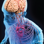
A study published in Nature offered insight into the way microglia may be playing a role in the central nervous system (CNS) symptoms of systemic lupus erythematosus (SLE).1 These brain cells are responsible for pruning and refining synapses, as well as cleaning up debris, such as amyloid plaque, but in disease, they may become inflamed or overprune.
Researchers looking at microglia in mice with lupus-like disease found that phagocytosis of neurons by microglia was increased in a way that was interferon-alpha-receptor dependent.
They also showed increased type 1 interferon signaling in the brains of deceased SLE patients.
“Under normal circumstances … microglia are phagocytosing synapses to keep them properly pruned,” Dr. Pope said. “Under the influence of type 1 interferon, you can see that there is increased uptake of this material. So one wonders: Could this be one of the mechanisms for CNS abnormalities in our patients with lupus—lupus fog and other symptoms that we see in our patients?”
In a study published in Nature Communications, researchers first showed that mice developed osteoarthritis when the adenosine receptor A2AR was knocked out. … The findings show potential for a new therapy.
Fli1 & Scleroderma
The transcription factor protein, Fli1, has been of interest in the scleroderma field for years, but in a study published in The Journal of Experimental Medicine, Fli1 was deleted only in epithelial cells.2
Researchers found that mice with keratinocytes in which Fli1 has been deleted develop dermal fibrosis, esophageal fibrosis and interstitial lung disease.
Dr. Pope said, “The really interesting part of this mouse model, to me, is the fact that these mice developed a systemic increase of IL-6 [interleukin 6] in the circulation,” along with antinuclear antibodies and autoantibodies to lung homogenates, which was not seen in control mice from the same litter.
Then researchers looked at medullary thymic epithelial cells (mTECs), which play an important role in self-tolerance. These cells are normally positive for Fli1 and autoimmune regulatory (Aire) proteins. But both are reduced in these cells in mice with Fli1 deleted from keratinocytes.


