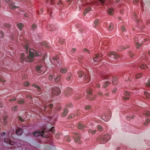
 SAN DIEGO—As part of a Nov. 14 session on lupus nephritis at ACR Convergence 2023, Simone Appenzeller, MD, PhD, shared perspectives on the importance of biopsy to inform its diagnosis, prognosis and treatment, with an emphasis on childhood disease.
Prompt diagnosis and treatment of lupus nephritis is perhaps even more important for children than for adults because of the risk of kidney failure and longer total lifetime impact. Kidney involvement in systemic lupus erythematosus is less common in adults than children, said Dr. Appenzeller, professor in the Department of Orthopedics, Rheumatology and Traumatology at the State University of Campinas, São Paulo, Brazil. She noted that kidney involvement is present in more than half of children with the disease. When lupus nephritis occurs in children, usually within the first two years of disease onset, it’s often more severe than in adults.1
Opinions on the optimal use of kidney biopsy in both adult and pediatric lupus have been evolving. Exactly when or how often the test should ideally be employed is unclear, and practitioners continue to weigh the diagnostic benefits of the gold standard test against the risks of an invasive procedure.2 Part of the diagnostic challenge is that established, non-invasive diagnostic markers often give incomplete information. For example, traditional laboratory markers, such as anti-double-stranded DNA antibody and complement, are often poor at detecting an early lupus nephritis flare; they also possess limited sensitivity and specificity in distinguishing active disease from earlier damage. Newer blood or urine markers and marker panels may eventually provide more informative results, but these are still likely to prove supplemental to biopsy.3

Dr. Appenzeller
The risk of kidney failure in childhood lupus is between 7% and 23%, added Dr. Appenzeller, and the mortality rate of pediatric patients with lupus nephritis is 19 times higher than those without nephritis.4
Renal Biopsy
Some patients with lupus nephritis have clear clinical or laboratory signs, such as edema or overt proteinuria, but others may have subtle or no symptoms. “We now know that clinical symptoms alone are not reliable enough to reflect the severity of renal disease in lupus nephritis; therefore, a renal biopsy is needed to guide treatment strategy,” said Dr. Appenzeller.
She highlighted the treat-to-target approach recently endorsed by the Pediatric Rheumatology European Society Taskforce, which strongly recommends early recognition and treatment of renal involvement, with a low threshold for renal biopsy to help reduce morbidity and mortality rates.5
Dr. Appenzeller also discussed a new clinical guideline from the nonprofit KDIGO (Kidney Disease Improving Global Outcomes). KDIGO recommends clinicians follow up abnormal urinalysis findings of proteinuria or urine sediment and proceed with biopsy if quantified proteinuria is greater than 0.5 g/day, or glomerular filtration rates are decreasing.6 For context, a 2012 clinical guideline from the ACR recommended kidney biopsy in patients with proteinuria >1.0 g/day or with proteinuria >0.5 g/day accompanied by hematuria or cellular casts.7 (Note: An updated ACR guideline on lupus nephritis is expected in 2025.)
Many clinicians opt not to perform a kidney biopsy if proteinuria doesn’t fall above a certain level. However, Dr. Appenzeller shared results from a cross-sectional study of 222 patients demonstrating that some important kidney pathology could be seen via histology in patients with lupus who had active urine sediment but low levels of proteinuria (<0.5 g/day). Thus, she recommended considering kidney biopsy even in patients with low-grade proteinuria.8
Histological Patterns
Lupus is a notoriously heterogenous disease, and lupus nephritis displays varying histological patterns as classified by the International Society of Nephrology/Renal Pathology Society (ISN/RPS). Patients with class I (minimal mesangial) and class II (mesangial proliferative) lupus nephritis typically have mild or absent clinical features. Patients with class III (focal disease) and class IV (diffuse disease) lupus nephritis have comparatively more acute injury, hematuria, proteinuria and chronic disease. Patients with class V (membranous disease) lupus nephritis have the highest rate of nephrotic syndrome, and class VI represents rare and advanced sclerosing disease.9
Dr. Appenzeller shared that biopsy patterns can also give clinicians helpful prognostic information. For example, a retrospective study of 382 patients with childhood lupus of class III or higher showed that patients with class III disease achieved complete remission more often than those with class IV or V disease.4
This same study highlighted the room for improvement in remission rates for childhood lupus: At 24-month follow-up, 57% of patients had achieved complete remission and 34% had achieved partial remission. Patients with severe disease at diagnosis had the highest risk of not achieving remission.4
Maintenance Therapy
Kidney biopsy can also enrich our understanding of true disease activity. For many years, researchers assumed that a complete clinical remission in lupus nephritis would be reflected in histological remission. But several studies have demonstrated this is not the case. Histologic findings may reveal ongoing inflammation not yet clinically apparent.10
“Repeat renal biopsy can help us identify complete remission more reliably,” explained Dr. Appenzeller. Because the optimal duration of maintenance immunosuppressive therapy for lupus nephritis is not clear, kidney biopsy can be an important tool in deciding whether to continue therapy. Biopsy helps clinicians balance the risks of ongoing immunosuppression vs. the risks of flare, which further increase the risks of chronic and end-stage kidney disease.10
Dr. Appenzeller discussed one prospective study of 75 patients during the maintenance phase of treatment for lupus nephritis. Roughly one-third of patients had histologic disease activity even though they had been in clinical remission for at least 12 months. The researchers continued maintenance immunosuppression in these patients, but allowed those with both ongoing clinical remission and histologic remission to stop their immunosuppression therapy. This approach seemed to decrease flare rates (1.5/year) compared with standard rates in the literature.10
Dr. Appenzeller also shared some complementary findings of a study showing histologic changes in repeated renal biopsies at diagnosis and consequent treatment of lupus nephritis. Higher inflammatorytype lesions—characterized by neutrophils, fibrinoid necrosis and cellular crescents—usually resolve completely during initial immunosuppression, she said. Standard-of-care treatment, including high-dose glucocorticoids, tends to work well for patients with such lesions.11
However, other lesions tend to persist with standard-of-care therapy, although at lower levels, in 50–60% of patients after eight to nine months of treatment. Many patients had other histological lesions even longer, especially hyaline deposits, glomerular crescents and tubulointerstitial inflammation. Such lesions often appear to take months or years to resolve, supporting the current use of long-term maintenance therapy in many patients.11
 Ruth Jessen Hickman, MD, a graduate of the Indiana University School of Medicine, is a medical and science writer in Bloomington, Ind.
References
- Groot N, de Graeff N, Avcin T, et al. European evidence-based recommendations for the diagnosis and treatment of childhood-onset lupus nephritis: The SHARE initiative. Ann Rheum Dis. 2017 Nov;76(12):1788–1796.
- Alforaih N, Whittall-Garcia L, Touma Z. A review of lupus nephritis. J Appl Lab Med. 2022 Oct;7(6):1450–1467.
- Palazzo L, Lindblom J, Mohan C, Parodis I. Current insights on biomarkers in lupus nephritis: A systematic review of the literature. J Clin Med. 2022 Sep;11(19):5759.
- De Mutiis C, Wenderfer SE, Basu B, et al. International cohort of 382 children with lupus nephritis—Presentation, treatment and outcome at 24 months. Pediatr Nephrol. 2023 Nov;38(11):3699–3709.
- Smith EMD, Aggarwal A, Ainsworth J, et al. Towards development of treat to target (T2T) in childhood-onset systemic lupus erythematosus: PReS-endorsed overarching principles and points-to-consider from an international task force. Ann Rheum Dis. 2023 Jun;82(6):788–798.
- Kidney Disease: Improving Global Outcomes (KDIGO) Lupus Nephritis Work Group. KDIGO 2024 clinical practice guideline for the management of lupus nephritis. Kidney Int. 2024 Jan;105(1S):S1–S69.
- Hahn BH, McMahon MA, Wilkinson A, et al. American College of Rheumatology guidelines for screening, treatment, and management of lupus nephritis. Arthritis Care Res (Hoboken). 2012 Jun;64(6):797–808.
- De Rosa M, Rocha AS, De Rosa G, et al. Low-grade proteinuria does not exclude significant kidney injury in lupus nephritis. Kidney Int Rep. 2020 Apr;5(7):1066–1068.
- Stokes MB, D’Agati VD. Classification of lupus nephritis; Time for a change? Adv Chronic Kidney Dis. 2019 Sep;26(5):323–329.
- Malvar A, Alberton V, Lococo B, et al. Kidney biopsy-based management of maintenance immunosuppression is safe and may ameliorate flare rate in lupus nephritis. Kidney Int. 2020 Jan;97(1):156–162.
- Malvar A, Alberton V, Lococo B, et al. Remission of lupus nephritis: The trajectory of histological response in successfully treated patients. Lupus Sci Med. 2023 May;10(1):e000932.

