Other well-recognized conditions that are occasionally associated with a positive ANA include chronic infectious diseases, such as mononucleosis, subacute bacterial endocarditis and tuberculosis; some lymphoproliferative diseases; and up to 90% of patients taking certain medications, especially procainamide and hydralazine, and those patients with rheumatoid arthritis treated with anti-TNF biologicals. However, most of the patients who take these medications and develop a positive ANA test do not develop clinical drug-induced lupus.
False-positive ANAs (i.e., ANAs in the absence of autoimmune disease or other diseases) are commonly found in normal women, elderly individuals and first-degree relatives of patients with ANA-positive autoimmune diseases (typically in low titer). There are several important points about ANA that should be considered in the clinical setting.
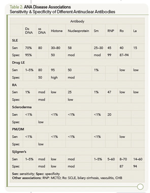
- The presence of high concentrations of antibody (titer >1:160) should make one suspicious that an autoimmune disorder is present. In this scenario, I recommend that sera then be tested for antibodies to double-stranded DNA (dsDNA), Sm, RNP, Ro (SS-A), La (SS-B) and, perhaps, Scl-70. The presence of antibodies to any of these greatly increases the likelihood that the patient has SLE, MCTD, Sjögren’s or scleroderma. Some labs will automatically test for these antibodies whenever the screening ANA is positive. However, the presence of these antibodies is not diagnostic of disease. If no initial diagnosis can be made, it is my practice to watch the patient carefully over time for the development of an ANA-associated disease and see the patient at least twice yearly.

- The combination of low titers of antibody (<1:80) and no or few signs or symptoms of disease portends a much smaller likelihood of an autoimmune disease developing. As a result, these patients with low ANA titers need to be reevaluated less frequently—yearly, unless clinical symptoms evolve to suggest an autoimmune disease.
- A patient with a negative ANA is highly unlikely to have either SLE, MCTD, Sjögren’s or scleroderma. However, if there is still strong clinical evidence of a systemic autoimmune disorder, one may test for the specific antibodies to double-stranded (ds) DNA, Sm, RNP, Ro, La or Scl-70, and if present increases the likelihood that they may have one of these autoimmune disorders. However, in my experience, these tests are typically negative. Nevertheless, it is prudent to see such patients where there is a high clinical index of suspicion, at least yearly—although more frequently if clinically indicated.
- Antinuclear antibodies produce a wide range of staining patterns (homogenous, diffuse, peripheral, rim, speckled, nucleolar, anticentromere, etc.). The nuclear staining pattern has been recognized to have a relatively low sensitivity and specificity for different autoimmune disorders. The presence of antibodies directed at specific nuclear antigens is usually more useful. (These antibodies include the following: dsDNA, Sm, RNP, Ro, La or Scl-70.)
Over the past few years, investigators and biotech firms have been developing solid-phase immunoassays to replace the IF ANA test.4-23 The rationale behind this attempt relates to performance characteristics of the IF technique. This test is very labor intensive and is subject to variation due to different interpretations by technicians. Also complicating testing is the fading of the image as it is examined in a fluorescent microscope. Furthermore, the IF technique uses serial dilutions of patient sera, which will give results that may not be linear. Variations in titer by twofold are common in day-to-day testing on the same sample; fourfold differences are said to be “significant.” By contrast, solid-phase immunoassays are automated and highly reproducible. The results are linear, and the technique is less labor intensive and, thus, cheaper to perform.
