Rheumatoid arthritis (RA) is characterized by polyarthritis, especially involving hands and wrists. Without treatment, RA usually evolves to articular deformities. Unfortunately, although rheumatoid deformities are characteristic, they are not pathognomonic, and we should be aware of possible mimics.1
Neuropathic arthropathy (NA), similar to other diseases, such as hemochromatosis, psoriatic arthritis, calcium pyrophosphate deposition disease, Jaccoud arthropathy, etc., is an example of an RA mimicker.2 This condition, also known as Charcot arthropathy, is marked by progressive joint dislocations and debilitating deformities. Any joint can be involved, depending on the underlying cause of the neuropathy. Although RA may involve the peripheral and central nervous system (mainly via compression, such as carpal tunnel syndrome and cervical myelopathy secondary to atlantoaxial subluxation), the association between RA and NA is so uncommon that every time we come across the possibility of it occurring, we must ask ourselves three questions:3-6
- Is my RA patient experiencing a complication of such a rare situation?
- Is my RA patient experiencing an overlap with another disease that promotes NA?
- Does my patient really have RA?
Case Report

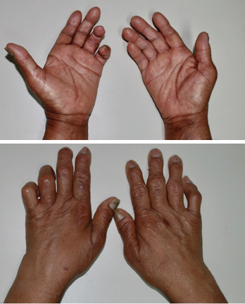
Figures 1 and 2: These images shows the patient’s bilateral ulnar deviation and benediction deformity of the left hand.
Consider the case of a 57-year-old female patient who presented to our rheumatology service to evaluate her for the introduction of biological therapy. She had been diagnosed with RA two years before. Her condition was refractory to methotrexate and leflunomide. Inflammatory markers, including erythrocyte sedimentation rate and C-reactive protein test, were mildly elevated, but she tested negative for rheumatoid factor (RF) and anti-citrullinated protein antibodies (ACPAs). Her complete blood count, electrolytes, urinalysis, renal and hepatic functions, and a viral panel were unremarkable.
The patient referred to mechanical hand pain and multiple unnoticed burn lesions on her upper extremities. Her physical examination was remarkable for benediction hands, ulnar deviation and interosseous muscle atrophy (see Figures 1 and 2), coupled with symmetric shoulder dislocations (see Figure 3, bottom right) and instability.
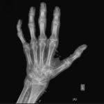
Her neurologic examination revealed dissociated sensory loss on her hands and arms, as well as limited shoulder abduction secondary to pain. She had already had a chest X-ray to screen for latent tuberculosis during the preparation to use biological therapy, and this radiography was remarkable for dislocation of both humeri (see Figure 4). Shoulder radiography evidenced incipient reabsorption of the ends of the humeri without prominent osteophytes or osteosclerosis characterizing the atrophic form of the Charcot joint (see Figure 5). Hand radiography, in addition to showing the deformities, was remarkable for destructive changes on her right wrist, as well as


