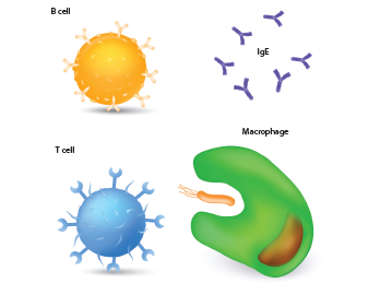
This figure illustrates immune system cells, including a macrophage, T cell, B cell and antibodies.
Designua / shutterstock.com
SAN DIEGO—B cell signaling goes awry in many patients with systemic lupus erythematosus (SLE), triggering pathogenic autoimmune responses and clinical disease. At the Rheumatology Research Foundation’s 2017 Evelyn V. Hess Memorial Lecture, held on Nov. 5 at the ACR/ARHP Annual Meeting, researcher Ignacio Sanz, MD, discussed B cells’ role in this complex disease.
Because lupus comprises multiple cell types during disease pathogenesis, and multiple mediators and signaling pathways that may contribute to disease, why do we need to emphasize B cells?
“Most of the disease risk alleles map to B cell pathways.1 SLE is characterized by multiple autoantibodies in very large amounts, and B cells also play autoantibody-independent functions that can be pathogenic,” including cytokine production, antigen presentation,
T cell activation and formation of tertiary lymphoid tissues, said Dr. Sanz, professor of medicine and pediatrics, chief of rheumatology and director of the Lowance Center for Human Immunology at Emory University, Atlanta. It is still unclear whether B cell therapies, such as rituximab, will be effective for some patient groups, such as African Americans with lupus nephritis.2
B cell depletion is highly variable in SLE, with two clear patterns: very profound depletion followed by repopulation with new B cells in patients who fare well long term, and “then another pattern [in which] patients might be declared complete B cell depletors by clinical standards, but in fact, they are not, and they tend to repopulate with memory cells and not do well,” said Dr. Sanz. These patients may not respond to such therapies as rituximab, he said. “You can deplete the B cells of a group of patients who are B cell dependent, and they will do well. The challenge is to recognize those patients and then, achieve that degree of depletion.”
Heterogeneous Disease
Researchers typically look at B cells in humans just by surface phenotypes or functional assays that scratch only the surface, said Dr. Sanz. To learn how B cells are programmed, or the mechanisms and directions they follow in the disease process, an immunomics approach works better.
Serum autoantibodies are analyzed in an SLE patient’s blood, kidney tissue, urine and bone marrow, then the researchers sequence the autoantibodies of most interest, including the immunoproteomics that help identify the dominant clones and B cell repertoire involved in disease activity. Through various sequencing techniques, the B cell repertoire is matched with the serum autoantibodies, “so we can understand the different profiles and classes of disease based on B cell signatures,” he said. “Then, we extract RNA and DNA from subsets of interests so we can study their molecular programs transcriptionally and epigenetically.”
At Dr. Sanz’s lab, this immunomics technique has been used to understand the role of plasmablasts in flaring SLE patients, for example.
Next, researchers isolate the B cells, and study the transcriptome and antibody repertoire, said Dr. Sanz. Conventional, six-color flow cytometry only shows a few naive B cell populations: memory cells, plasmablasts or double-negative (DN) cells. When he and his young sons decorated a sidewalk with colored chalk, he realized more colors were necessary. By using 12-color cytometry, “we can now identify up to 29 populations of B cells that are very reproducible and discrete, and you can identify how they may be dysregulated in SLE,” he said. Double-negative B cells, including DN1 and DN2, are intriguing in lupus pathogenesis. “Critical insights can come from unexpected sources.”
‘You can deplete the B cells of a group of patients who are B cell dependent, & they will do well. The challenge is to recognize those patients & then achieve that degree of depletion.’ —Ignacio Sanz, MD
Dr. Sanz and his team use 12-color cytometry in an ongoing study of 135 SLE patients whose B cell profiles are compared to 25 healthy controls. Profiles of one group of SLE patients, those with a loss of unswitched memory cells and expansion of newly defined activated naive cells and DN2 cells, look very different from other lupus patients with B cell profiles quite similar to those of healthy individuals. The first subset of patients tend to have higher disease activity and include a high concentration of African Americans, more nephritis and skin disease, high anti-Smith, high anti-RNP and high interferon-α, he said. The profiles are then mapped to measure the distance from a mean of normal B cell activity in healthy people. Patients who map farthest from the normal mean flare faster, more often and more intensely, he said.
Dr. Sanz said, “We believe this means there’s probably a B cell-dependent disease and a B cell-independent disease.” Patients with dependent disease may flare more severely and respond better to B cell therapies, but the opposite is true for those with independent disease. Using this mapping exercise, they also explored B cell abnormalities in chronic cutaneous lupus and explored patients’ risk for progression to SLE.3
Immunomics may help identify specific molecular targets or pathways for subsets of SLE patients that lead to innovative clinical trials with mechanistic endpoints that reveal both responders to treatment and nonresponders, who are just as important to understand, said Dr. Sanz.
Double-Negative Cells
The B cell pathway may be split into two compartments: germinal cell-derived with long-lived memory cells and plasma cells, and then an extrafollicular pathway that derives from new activation of autoreactive, naive cells that activate and generate short-lived plasmablasts, he said. This second pathway is very active in lupus flares. Double-negative B cells are highly prevalent in the peripheral blood in some patients, mostly African Americans with active lupus, and usually coincide with large expansions of plasmablasts. Lupus patients with a large expansion of DN2 cells also have high levels of CD19, and lack the follicular marker CXCR5. DN2 cells are also prominent in cohorts of African American SLE patients.
“It’s also been proposed that DN2 cells are associated with age. We don’t find that age association in SLE. In lupus, it’s very striking that even in very young patients with acute disease onset, essentially all the B cells in the blood are already DN2s,” he said. They also correlate with high levels of anti-Smith autoantibody and high SLEDAI (a measure of SLE disease activity). Molecular programming of DN2 B cells in lupus patients points to one marker, SLAMF7, which may have therapeutic implications. Elotuzumab, a SLAMF7 inhibitor, is already approved by the U.S. Food and Drug Administration (FDA) for treatment of refractory multiple myeloma.
DN2 cells are also highly responsive to interferon-λ, which is another therapeutic target of growing interest in lupus research. It is also possible that an anti-SLAMF7 antibody therapy might be able to wipe out both the extrafollicular B cell pathway and pre-established, long-standing plasma cells, which Dr. Sanz’ group are studying in a current clinical trial.
Epigenetics
Epigenetic dysregulation of B cells in SLE provides more clues. Conventional studies of B cells are too heterogeneous, so results can be misleading, said Dr. Sanz. At his laboratory, RNA sequencing, bisulfite sequencing and ATAC sequencing is used to understand DNA methylation, chromatin accessibility and their impact on transcriptional activity of subsets of B cells, generating a large data set. They hope to discover whether epigenetic programs can identify distinct fates, activation and differentiation of particular B cell pathways, and if SLE-specific B cell programs can help segment disease populations to identify more precise therapies, serving as surrogates for disease developments, remission, therapeutic response and B cell tolerance, he said.
“If we could at least show that disease-related, epigenetic modifications are reversed, it would be a very good surrogate of the disease response and, perhaps, B cell tolerance,” he said. His current research in this area includes methylation studies, which reveal significant clues. “There are a few thousand loci in lupus B cells that are more methylated than in healthy controls, and with RNA sequencing, we find that 1,000 more are highly expressed in lupus B cells and 500 are down-regulated.”
Epigenetic research in SLE shows that resting naive B cells display a poised pathogenic signature even before activation and have a unique chromatin accessibility signature, he said.4 Mounting evidence suggests that dysregulation of lupus B cells starts very early in the disease process in the patient’s bone marrow.
Therapeutic Horizons
B cell research may lead to new, more effective therapies for specific subsets of SLE patients. These include histone deacetylase inhibitors, which may modify renal disease by regulating B cells epigenetically to modulate their response in lupus nephritis patients, he said.
Patients with SLE produce autoantibodies for years before clinical disease appears and progress through normal immunity to benign immunity to pathogenic autoimmunity and, finally, to clinical disease. Even young children with pediatric SLE may produce antinuclear autoantibodies but have a normal B cell profile and no clinical disease, he said. This can progress rapidly to a dysregulated B cell profile and, presumably, epigenetic marking that could allow those patients to be targeted for particular B cell therapies, he said. Chimeric antigen receptor (CAR) T cell-based therapies are another intriguing area of lupus research.5
“I don’t think we are yet at the point to use an antigen-specific approach in lupus, but I think the CAR T cells, particularly C-19 CAR T cells, that deplete B cells and a major fraction of positive C-19 plasma cells, could be used for that desired, persistent, profound B cell depletion.”
Susan Bernstein is a freelance journalist based in Atlanta.
References
- Mohan C, Putterman C. Genetics and pathogenesis of systemic lupus erythematosus and lupus nephritis. Nat Rev Nephrol. 2015 Jun;11(6):329–341.
- Rovin BH, Furie R, Latinis K, et al. Efficacy and safety of rituximab in patients with active proliferative lupus nephritis: The Lupus Nephritis Assessment with Rituximab study. Arthritis Rheum. 2012 Apr;64(4):1215–1226.
- Wei C, Hill A, Smith K, et al. B cell abnormalities in patients with chronic cutaneous lupus erythematosus [abstract]. Arthritis Rheumatol. 2017;69(suppl. 10).
- Scharer CD, Blalock EL, Barwick BG, et al. ATAC-seq on biobanked specimens defines a unique chromatin accessibility structure in naive SLE B cells. Sci Rep. 2016 Jun 1;2016(6):27030.
- Ellebrecht CT, Bhoi VG, Nace A, et al. Re-engineering chimeric antigen receptor T cells for targeted therapy of autoimmune disease. Science. 2016 Jul;353(6295):179–184.
